

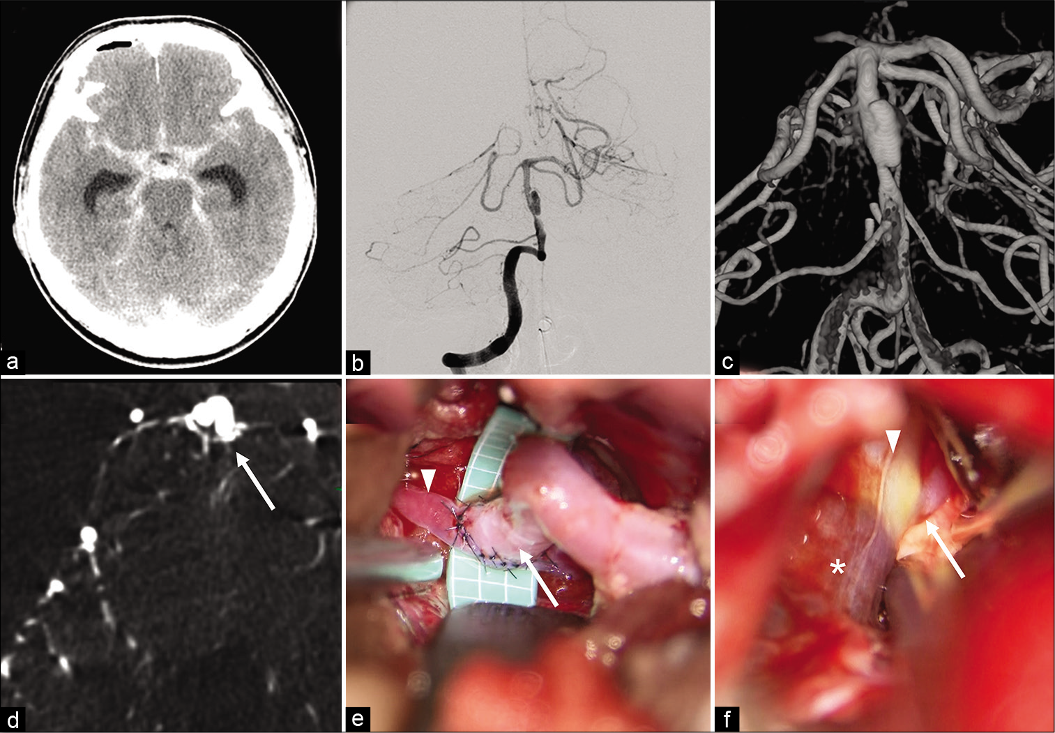
Superficial temporal artery-superior cerebellar artery bypass and proximal occlusion through anterior petrosal approach for subarachnoid hemorrhage due to basilar artery dissection
Date of publication: 21-Aug-2020
Background: Subarachnoid hemorrhage (SAH) due to rupture of basilar artery dissection (BAD) is extremely rare and often has a poor prognosis. Since ruptured BAD has high rate of rebleeding and mortality, treatment to prevent rerupture is mandatory in the acute phase. However, to date, no optimal treatment has been established which satisfies secure prevention of rerupture and ischemia simultaneously. Herein, we report a case of SAH due to BAD treated with proximal occlusion of basilar artery with superficial temporal artery (STA)-superior cerebellar artery (SCA) bypass, preventing rebleeding securely and ensuring adequate blood flow in the upper basilar region.
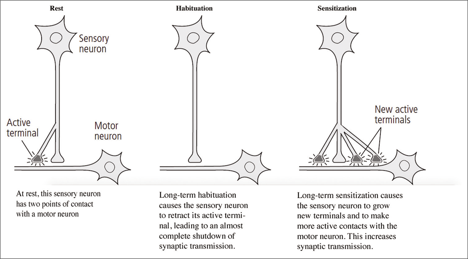
The neurobiology of learning and memory – as related in the memoirs of Eric R. Kandel
Date of publication: 15-Aug-2020
Comment regarding the article “Posttraumatic thoracic epidural capillary hemangioma”
Date of publication: 15-Aug-2020
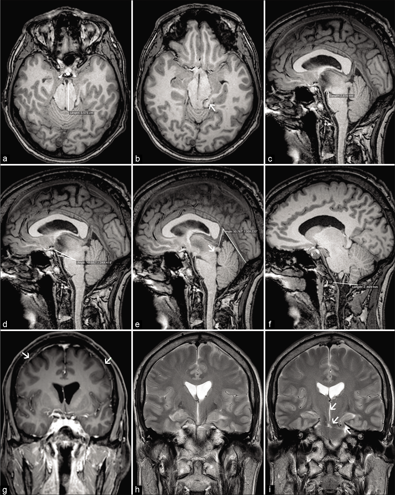
Frontotemporal brain sagging syndrome: Craniospinal hypovolemia secondary to a T6-T7 cerebrospinal fluid-venous fistula
Date of publication: 15-Aug-2020
Background: The frontotemporal brain sagging syndrome (FTBSS) is defined as an insidious/progressive decline in behavior and executive functions, hypersomnolence, and orthostatic headaches attributed to cerebrospinal fluid (CSF) hypovolemia. Here, a T6 CSF-venous fistula (e.g., between the subarachnoid CSF and a paraspinal vein) resulted in a CSF leak responsible for craniospinal hypovolemia.
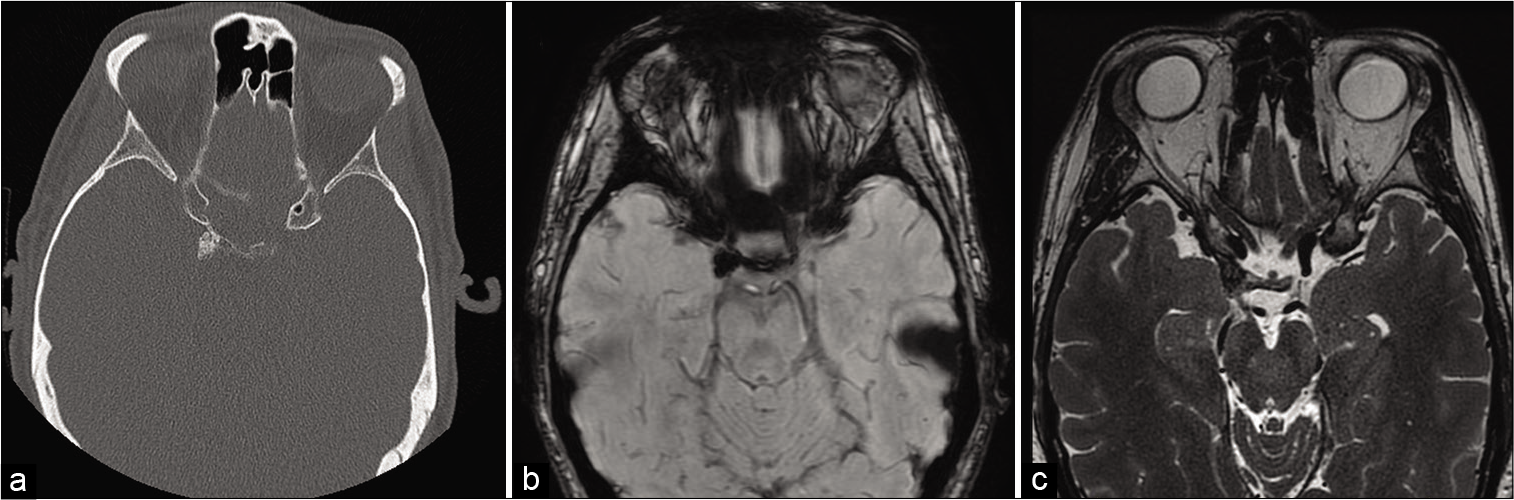
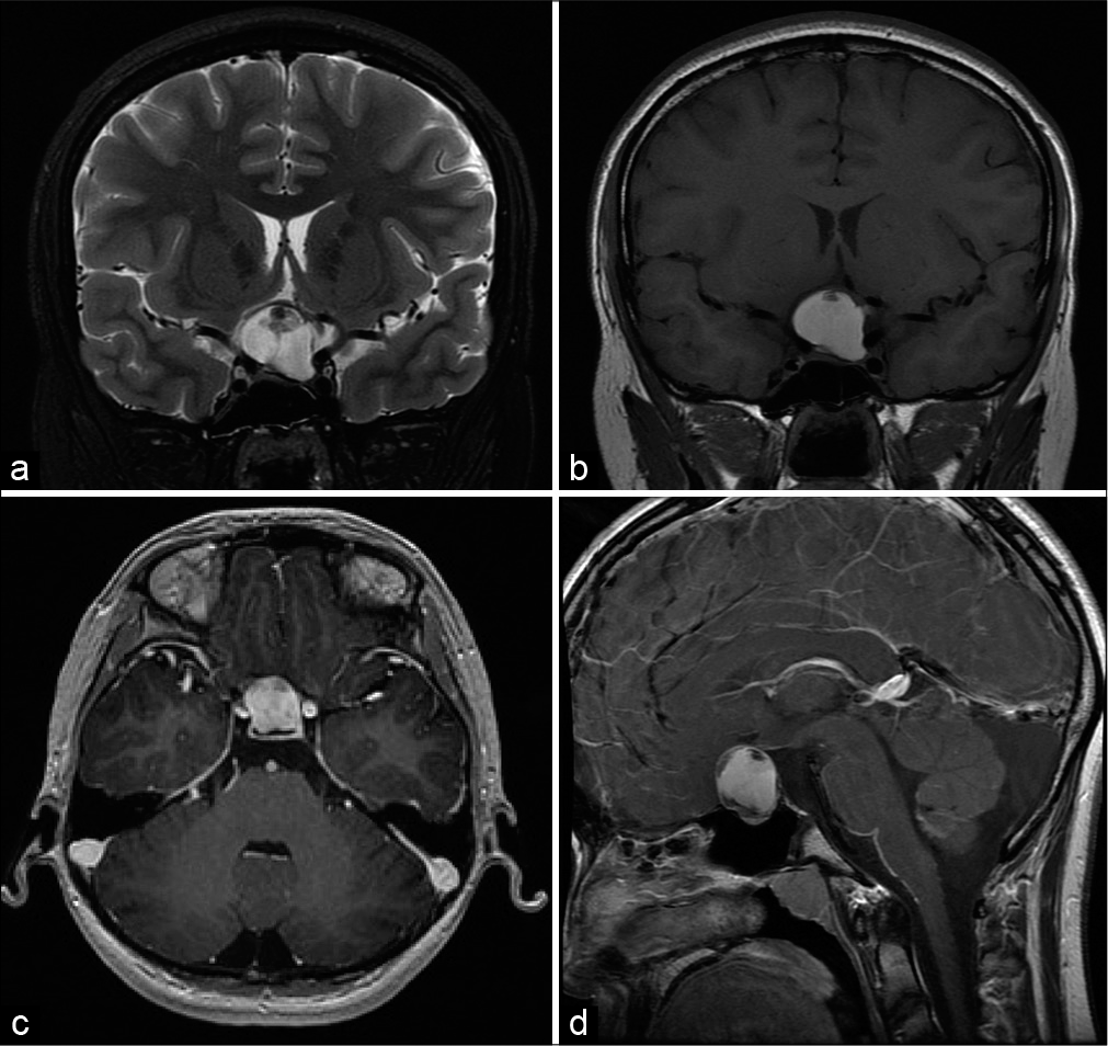
Calcifying pseudoneoplasm of the neuraxis: A rare case involving the oculomotor nerve
Date of publication: 15-Aug-2020
Background: Calcifying pseudoneoplasm of the neuraxis (CAPNON) is a rare entity which can occur at intracranial and spinal locations. Clinical presentation is due to local mass effect rather than tissue infiltration. Lesions causing significant symptoms or are showing radiological progression require surgical resection. Maximal surgical resection is considered curative for this non-neoplastic entity with only two cases of recurrence reported in the literature. Cranial nerve involvement is extremely rare and the presenting neurological deficit is unlikely to improve even with surgical intervention.
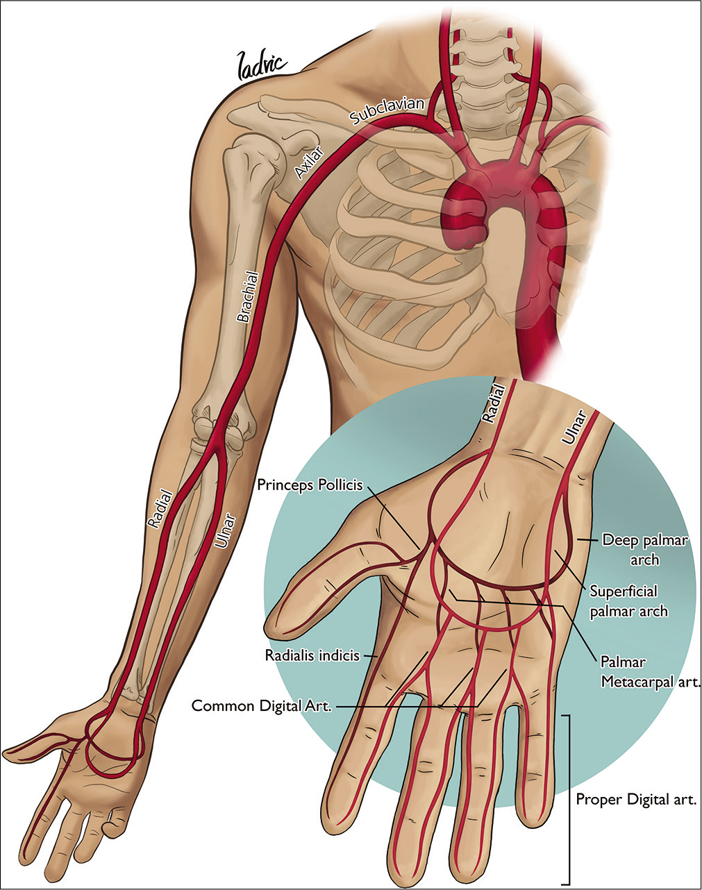
Transradial approach and its variations for neurointerventional procedures: Literature review
Date of publication: 15-Aug-2020
Background: The transfemoral approach (TFA) has been the standard in neuroradiology over the years. However, the transradial approach (TRA) and its variants offer several benefits over the TFA.
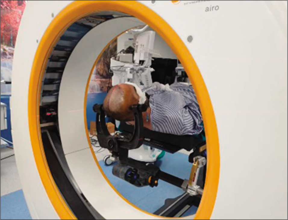
Role of intraoperative computed tomography scanner in modern neurosurgery – An early experience
Date of publication: 15-Aug-2020
Background: Intraoperative imaging addresses the limitations of frameless neuronavigation systems by providing real-time image updates. With the advent of new multidetector intraoperative computed tomography (CT), soft tissue can be visualized far better than before. We report the early departmental experience of our intraoperative CT scanner’s use in a wide range of technically challenging neurosurgical cases.
Rathke’s cleft cyst with xanthogranulomatous change: A case report and review of the literature
Date of publication: 15-Aug-2020
Background: Rathke’s cleft cysts (RCCs) are benign, typically asymptomatic sellar lesions found incidentally in adults, with a dramatically lower incidence in pediatric patients (<18 years). We present a case of RCC with xanthogranulomatous change (XGC) – an even less common subtype of RCC – treated by endoscopic endonasal surgical resection. This is the second reported instance of an RCC with XGC occurring in a pediatric patient.
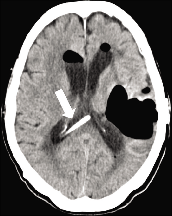
Spontaneous cerebrospinal fluid otorrhea and pneumocephalus on the contralateral side of the previous cranial surgery
Date of publication: 15-Aug-2020
Background: Cerebrospinal fluid (CSF) leaks and pneumocephalus commonly occur due to head trauma or surgical procedures. Spontaneous CSF (sCSF) leaks, however, occur without any clear etiology and are relatively uncommon.
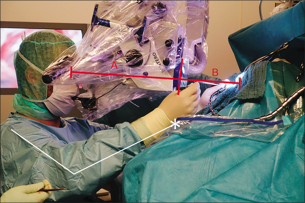
Ergonomics of surgical microscopes for the sitting position as determined by ocular-corpus length
Date of publication: 15-Aug-2020
Background: The sitting position is favorable for microsurgical procedures applied to posterior midline pathologies in both the supra- and infratentorial regions. The dimensions of the microscope corpus affect the device’s comfort and handling in the hands of the microneurosurgeon for such procedures. A shorter microscope corpus provides more favorable intraoperative ergonomics for surgical practice.

