

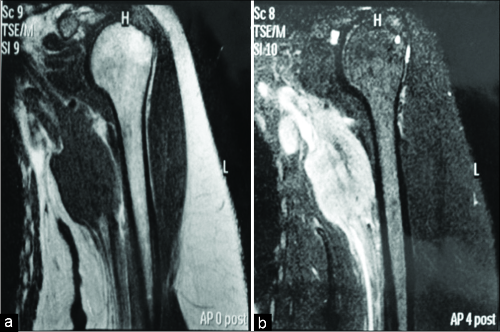
Multimodal treatment of an extremely rare desmoplastic small round cell tumor primary to the brachial plexus – A case report and review of literature
Date of publication: 15-Jul-2019
Background: Desmoplastic small round cell tumor (DSRCT) is a rare and aggressive malignant neoplasm typically located in the abdomen or pelvis. Other possible locations are the chest, pleura, scrotum, and central nervous system. DSRCT originally arising from the brachial plexus (BP) is extremely rare, to the best of our knowledge, only two cases have been previously described in the English scientific literature.
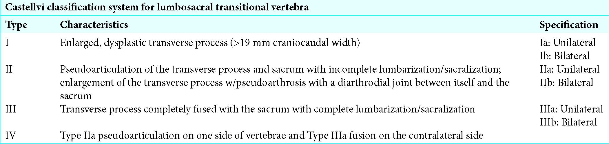
Surgical management of Bertolotti’s syndrome in two adolescents and literature review
Date of publication: 05-Jul-2019
Background: Bertolotti’s syndrome is defined by back pain and/or radicular symptoms attributed to a congenital lumbosacral transitional vertebra (LSTV). There are few studies that discuss the surgical management of Bertolotti’s syndrome. Here, we report long-term outcomes after resecting a pseudoarthrosis between the sacrum and L5 in two teenage patients, along with a review of literature.
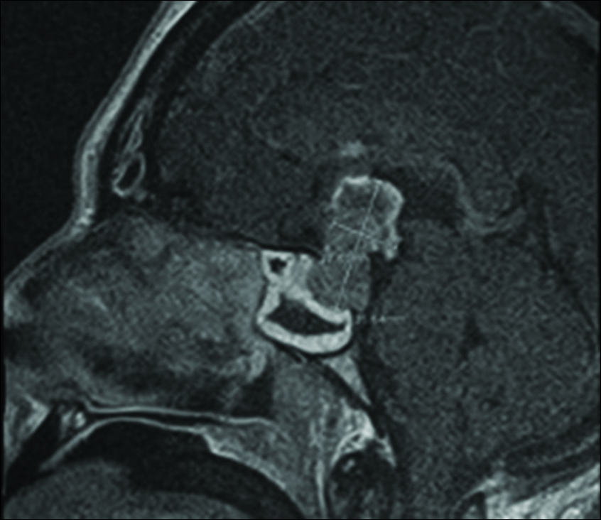
Atypical teratoid/rhabdoid tumor presenting with subarachnoid and intraventricular hemorrhage
Date of publication: 05-Jul-2019
Background: Sellar masses comprise 14–18% of all intracranial tumors. Pituitary adenomas account for 85% of these lesions, while 15% of sellar masses stem from other etiologies. Intratumoral hemorrhage (apoplexy), while not exceptionally common, can be discovered at presentation. While the hemorrhage pattern is typically contained within the tumor, an extension of bleeding beyond the sella has been reported.
Minimally invasive posterior fossa decompression in Chiari I malformation
Date of publication: 05-Jul-2019
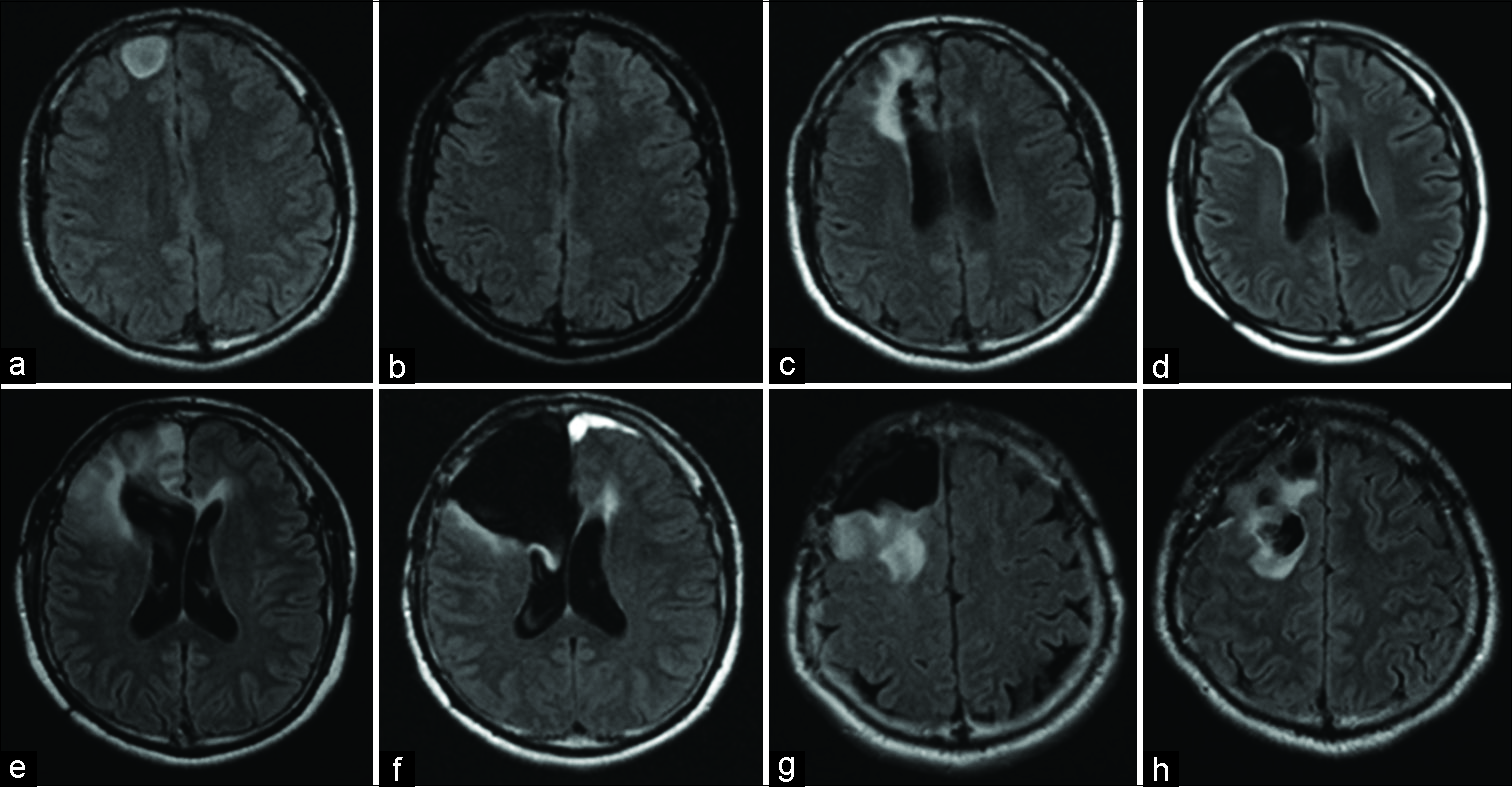
An autopsy case of widespread brain dissemination of glioblastoma unnoticed by magnetic resonance imaging after treatment with bevacizumab
Date of publication: 05-Jul-2019
Background: Although glioblastoma has been shown to be able to disseminate widely in the intracranially after treatment with bevacizumab without any significant radiological findings, reports on such cases with subsequent autopsy findings are lacking.
Understanding gamma ventral capsulotomy: Potential implications of diffusion tensor image tractography on target selectivity
Date of publication: 05-Jul-2019
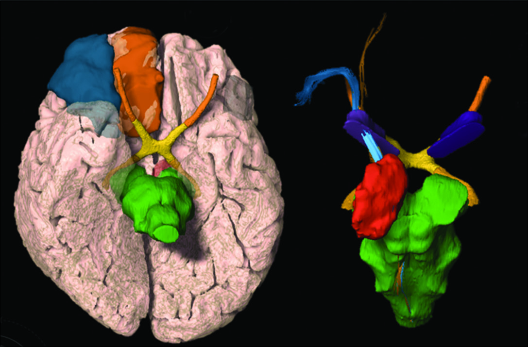
Background: The role of tractography in gamma ventral capsulotomy (GVC) planning is still unclear. This paper aims to describe the spatial distribution of medial orbitofrontal cortex (OFC) and lateral OFC fibers passing through the anterior limb of the internal capsule (ALIC) and analyze quantitative tractography parameters that differentiate obsessive-compulsive disorder (OCD) individuals from other neurosurgery functional patients (morbid obesity and Parkinson’s disease [PD]).

Surgical management of Bertolotti’s syndrome in two adolescents and literature review
Date of publication: 05-Jul-2019
Background: Bertolotti’s syndrome is defined by back pain and/or radicular symptoms attributed to a congenital lumbosacral transitional vertebra (LSTV). There are few studies that discuss the surgical management of Bertolotti’s syndrome. Here, we report long-term outcomes after resecting a pseudoarthrosis between the sacrum and L5 in two teenage patients, along with a review of literature.
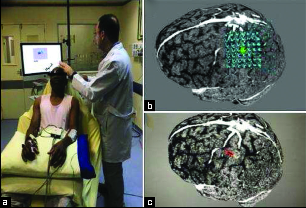
Computed tomography-guided navigated transcranial magnetic stimulation for preoperative brain motor mapping in brain lesion resection: A case report
Date of publication: 05-Jul-2019
Background: Navigated transcranial magnetic stimulation (nTMS) is a well establish a noninvasive method for preoperative brain motor mapping. We commonly use magnetic resonance imaging (MRI) to supply the nTMS system. In some cases, MRI is not possible or available, and the use of computed tomography (CT) is necessary. We present the first report describing the association of CT and nTMS motor mapping for brain lesion resection.
Unedited microneurosurgery of a falcotentorial meningioma
Date of publication: 05-Jul-2019
Background: Falcotentorial meningiomas are pineal region meningiomas that arise from the dura of the tentorium cerebelli and posterior part of the falx. These tumors are commonly supplied by branches of the internal carotid artery such as the meningohypophyseal trunk, inferolateral trunk, and anterior choroidal artery. Less frequently, branches of the ophthalmic artery, vertebral artery, or external carotid artery are also involved. Based on neuroimaging studies, falcotentorial meningiomas may be classified as anterior, superior, inferior, and posterior types. Here, we present an unedited microsurgical resection of a superior falcotentorial meningioma.
Potential improvement of survival statistics for glioblastoma multiforme (WHO IV)
Date of publication: 28-Jun-2019
Abstract
The present-day treatment of a glioblastoma multiforme IV (glioblast) is by surgery, radiation, and chemotherapy. Unfortunately, the current treatment has not significantly improved the survival statistics of this tumor. There are now two relatively new surgical procedures that may improve the survival statistics of this malignancy. One of these procedures is the intraoperative use of the drug 5-aminovolumic acid (ALA), which fluoresces a red color in malignant brain tissue that is not observed in normal brain tissue. This allows a neurosurgeon to distinguish brain tissue infiltrated by malignant cells, thus allowing a more complete resection of the tumor. Another procedure that has the potential to improve the survival statistics of glioblasts is the use of the omentum. Direct placement of the omentum on a brain infiltrated by malignant cells would allow omental blood vessels, known to be completely clear of endothelial cells, to penetrate directly into the underlying brain. The blood flow through omental blood vessels could be expected to carry chemotherapeutic agents throughout the involved brain, thereby totally bypassing the blood–brain barrier. Combining a tumor resection using 5-ALA and placing the omentum on the brain may prove instrumental in improving the survival statistics of patients suffering from a glioblast.

