

Multiple meningiomas arising within the same hemisphere associated with Li-Fraumeni syndrome
Date of publication: 17-Mar-2021
Background: While meningiomas are some of the most common intracranial tumors, the presence of multiple ones at the time of presentation is rare and can most commonly be observed in patients with well-described syndromes (i.e., neurofibromatosis type 2) or those with prior cranial radiation history. In others, however, the pathophysiology remains unclear.
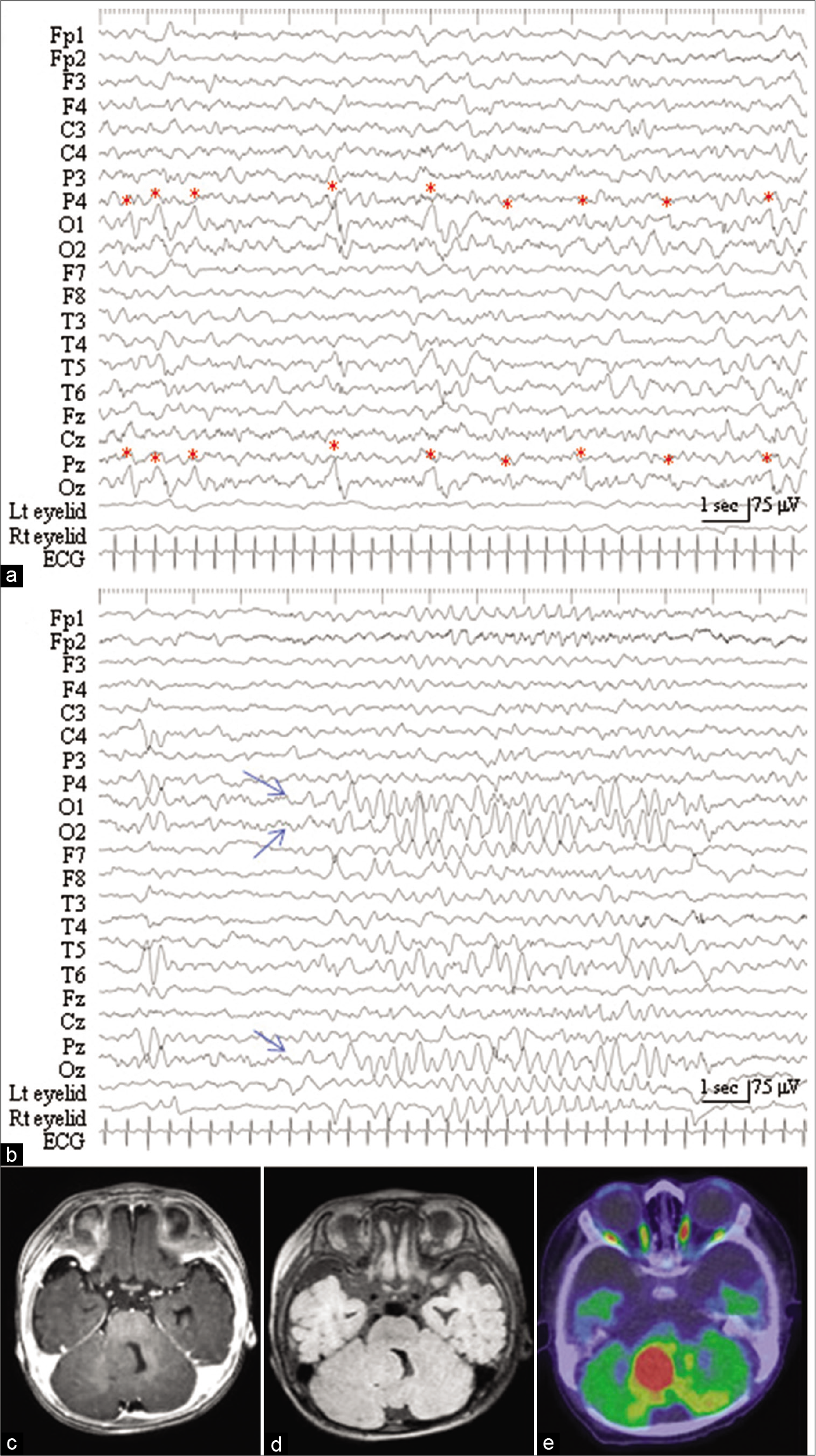
Periodic discharges with high frequency oscillations recorded from a cerebellar gangliocytoma in an epileptic infant
Date of publication: 17-Mar-2021
Background: Subcortical epilepsies associated with developmental tumors in the cerebellum are rarely experienced. As supportive evidence of the intrinsic epileptogenicity of cerebellar tumors, previous electroencephalogram (EEG) studies with intratumoral depth electrodes demonstrated epileptiform or ictal discharges. Recent studies have demonstrated that high frequency oscillations (HFOs) can be regarded as a new biomarker of epileptogenesis and ictogenesis; however, there are few evidence about HFOs in cases of epilepsy associated with cerebellar tumors.
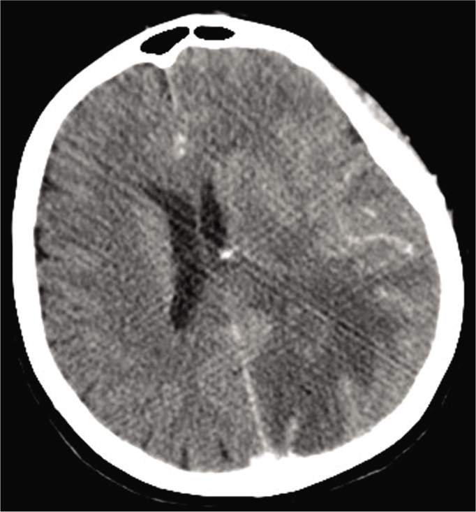
Brain abscess in a rheumatoid arthritis patient treated with leflunomide – A case presentation and review
Date of publication: 17-Mar-2021
Background: Immunosuppression is a significant parameter in the pathogenesis of brain abscesses (BA) and it could be the result of severe infections such as acquired immunodeficiency syndrome or drug-induced, by several medications used for systemic autoimmune diseases. Leflunomide is a pyrimidine synthesis inhibitor that affects the proliferation of lymphocytes and is used as a disease-modifying antirheumatic drug. Mild infections, particularly those of the respiratory tract and herpes zoster, are one of its most common adverse effects. However, atypical and severe infections have also been reported under treatment with leflunomide.
Transsulcal parafascicular brain path-assisted approach to subcortical lesions: 2-dimensional operative video
Date of publication: 17-Mar-2021
Background: Approaches to subcortical lesions have traditionally been limited by the morbidity of white matter dissection and fixed blade retraction required to reach these targets. Visualization of deep surgical fields with a traditional operating microscope is also poor. Coordinated use of intra-operative image guidance, a tubular retractor (BrainPath®, Nico Corp, Indianapolis, Indiana), a high-definition exoscope (Vitom®, Karl Storz Endoscopy America, Inc, El Segundo, California), and a low-profile resection device (Myriad®, Nico Corp) facilitates atraumatic access to and resection of subcortical lesions including primary brain tumors, brain metastases, and intracerebral hemorrhages.[
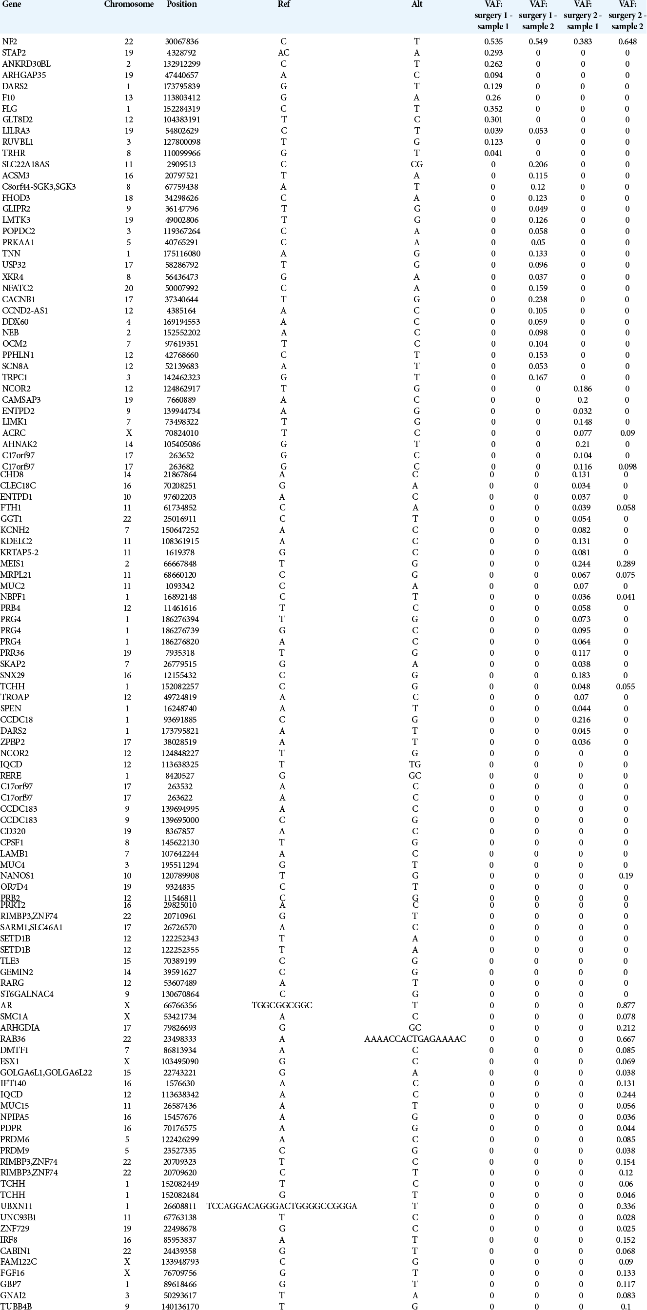
Erratum: Profile of genetic variations in severely calcified carotid plaques by whole-exome sequencing
Date of publication: 13-Mar-2021
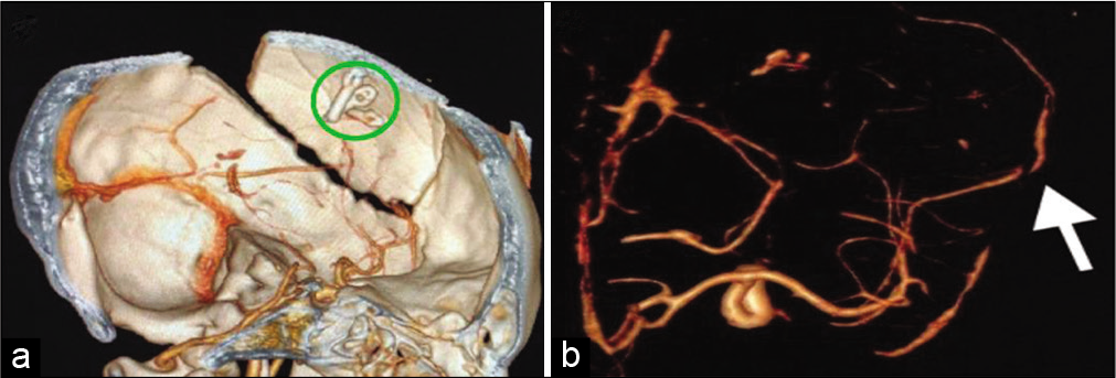
Ruptured giant aneurysm of a cortical middle cerebral artery: A case report
Date of publication: 08-Mar-2021
Background: Aneurysms of the cortical branches of the middle cerebral artery (MCA) are rare. They usually are secondary to traumatic or infectious etiologies and are rarely idiopathic. The specific characteristics of idiopathic aneurysms in such location are not well defined in the literature. The authors report a rare case of a ruptured giant idiopathic cortical MCA aneurysm with review of the available literature on this clinical entity.
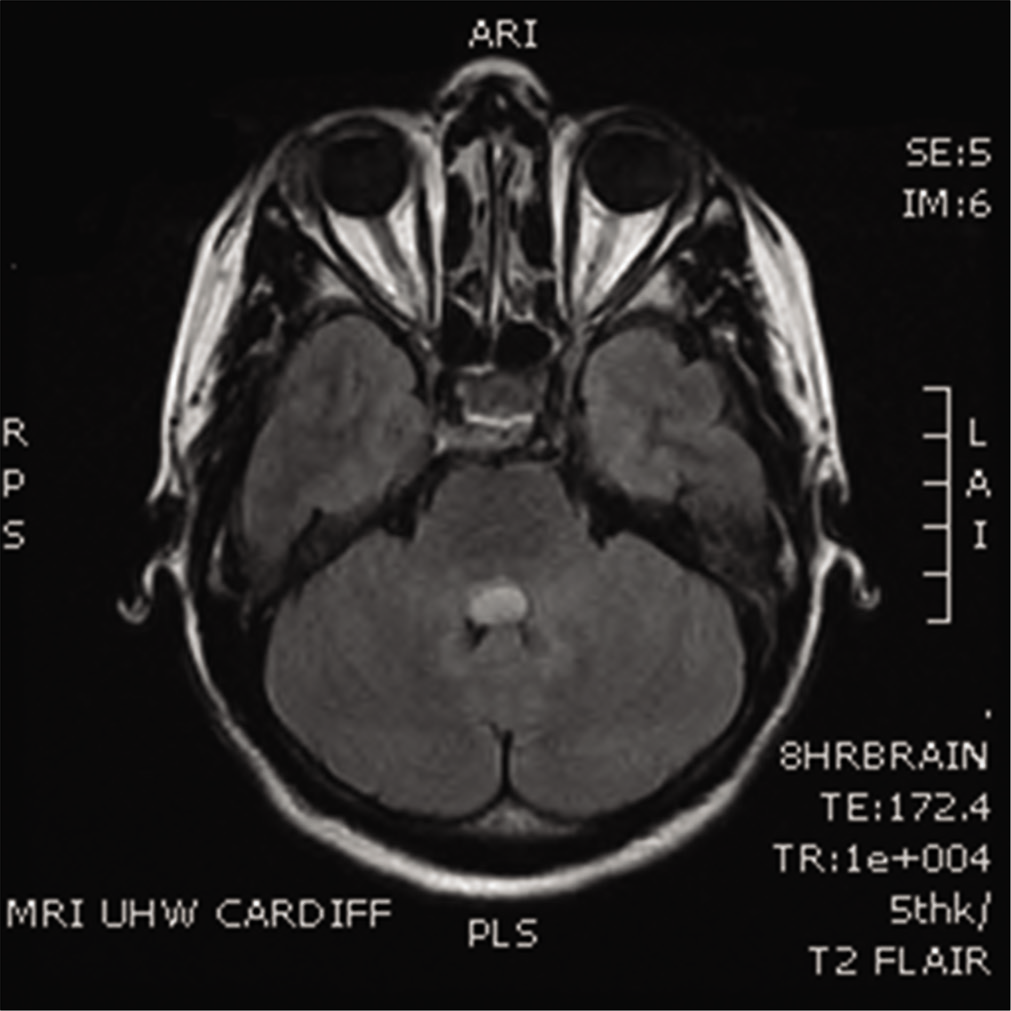
Mollaret’s triangle: An important neuroanatomical territory for all clinicians
Date of publication: 08-Mar-2021
Background: Hypertrophic olivary degeneration is a rare condition caused by damage within the triangle of Guillain and Mollaret. We discuss the anatomical, radiological, and clinical history of this rare condition.
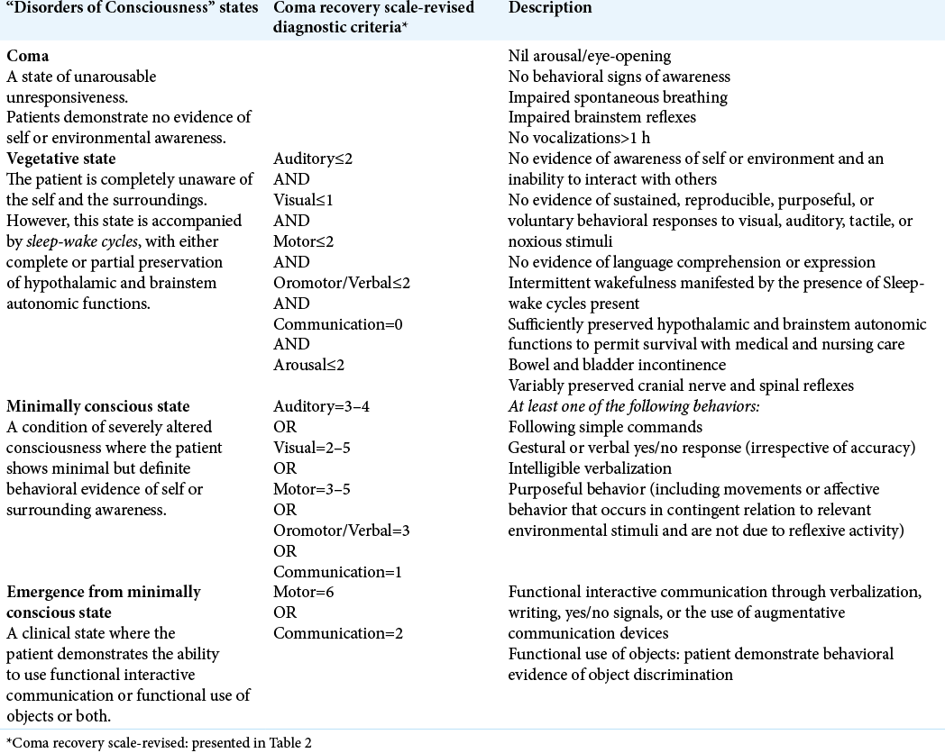
Coma recovery scale: Key clinical tool ignored enough in disorders of consciousness
Date of publication: 08-Mar-2021
Background: Disorders of consciousness (DoC) includes coma, vegetative state (VS), minimally conscious state (MCS), and emergence from the MCS. Aneurysmal rupture with high-grade SAH, traumatic brain injury, and neoplastic brain lesions are some of the frequent pathologies leading to DoC. The diagnostic errors among these DoC are as high as ranging from 25% to 45%, with a probable error in the conclusion of patients’ state, treatment choice, end-of-life decision-making, and prognosis. Some studies also reported that 37–43% of patients were misdiagnosed in VS while demonstrating signs of awareness. Despite its wide acceptance, Coma Recovery Scale-Revised (CRS-r) remained underused or inappropriately utilized, which may lead to substandard or unprofessional patient care. Literature is rare on the knowledge of CRS-r among physicians published from India and across the globe. Therefore, we carried out the present study to ascertain physicians’ knowledge on CRS-r and raise awareness about its justifiable clinical utilization. We also explored the factors associated with this perceived level of experience among participants and recommend frequent physicians’ training for care of patients with DoC.
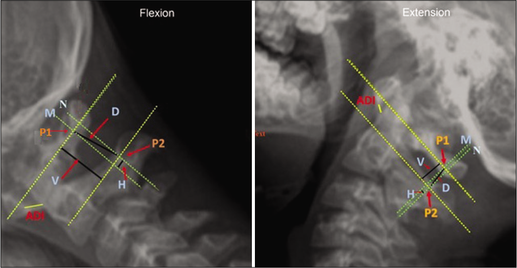
Fiberoptic bronchoscopy versus video laryngoscopy guided intubation in patients with craniovertebral junction instability: A cinefluroscopic comparison
Date of publication: 08-Mar-2021
Background: Manipulation during endotracheal intubation in patients with craniovertebral junction (CVJ) anomalies may cause neurological deterioration due to underlying instability. Fiberoptic-bronchoscopy (FOB) is better than video laryngoscope (VL) for minimizing cervical spine movement during intubation. However, evidence suggesting superiority of FOB in patients with CVJ instability is lacking. We prospectively compared dynamic movements of the upper cervical spine during intubation using FOB with VL in patients with CVJ anomalies.
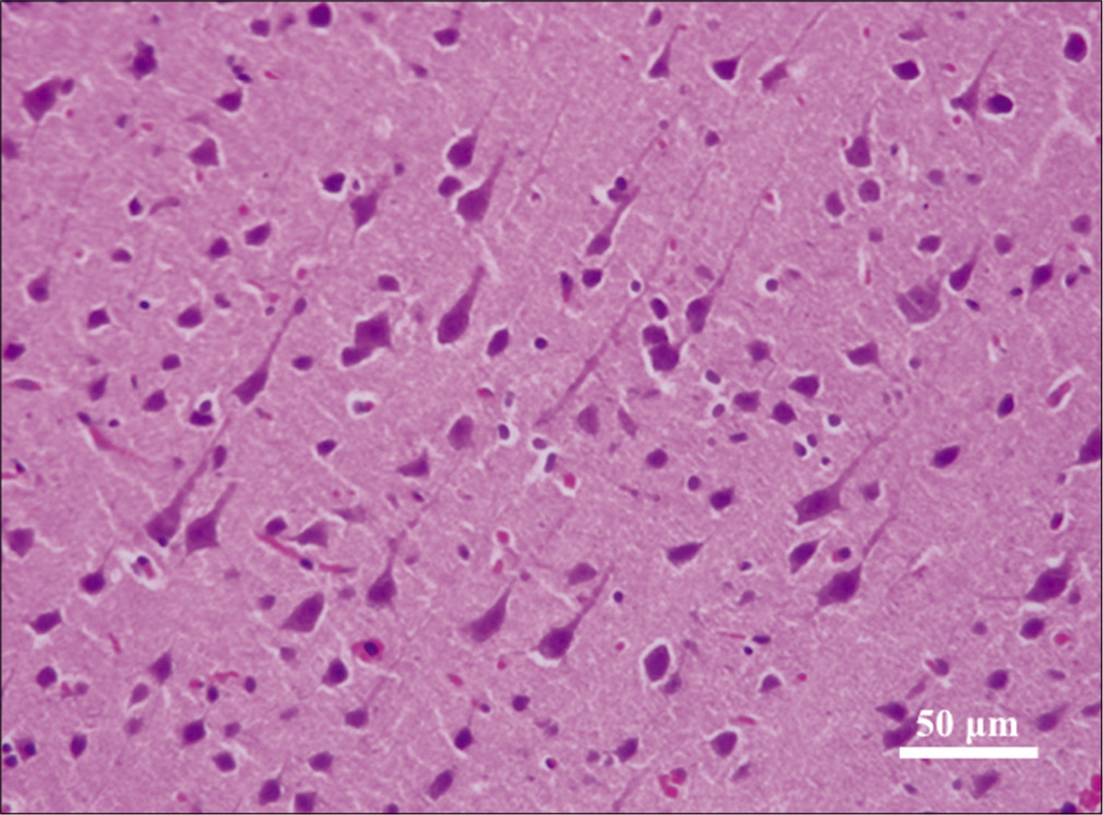
Histopathological changes of neuronal tissue following the use of hydrogen peroxide in neurosurgical procedures
Date of publication: 08-Mar-2021
Background: Hydrogen peroxide (HP) is routinely used in neurosurgical procedures to achieve surgical hemostasis. However, its safety profile is still debatable with various reports depicting range of adverse effects on neuronal tissue. The objective of this paper is to evaluate the safety and efficacy of HP as a hemostatic agent in normal neuronal tissue during neurosurgical procedures conducted on rats.

