

Intraparenchymal subependymoma: Case report and literature review
Date of publication: 14-Apr-2021
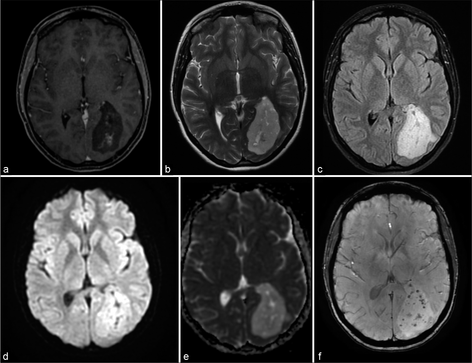
Background: Intracranial subependymomas are rare slow-growing benign tumors typically located in the ventricular system, accounting for 0.07–0.7% of all intracranial neoplasms. Intraparenchymal subependymoma is extremely rare lesions, imposing a challenging diagnosis and management.
Osteosarcoma of the temporal bone occurring 40 years after radiotherapy: A technical case report presenting en bloc resection of intra- and extracranial lesions followed by a one-stage reconstruction
Date of publication: 14-Apr-2021
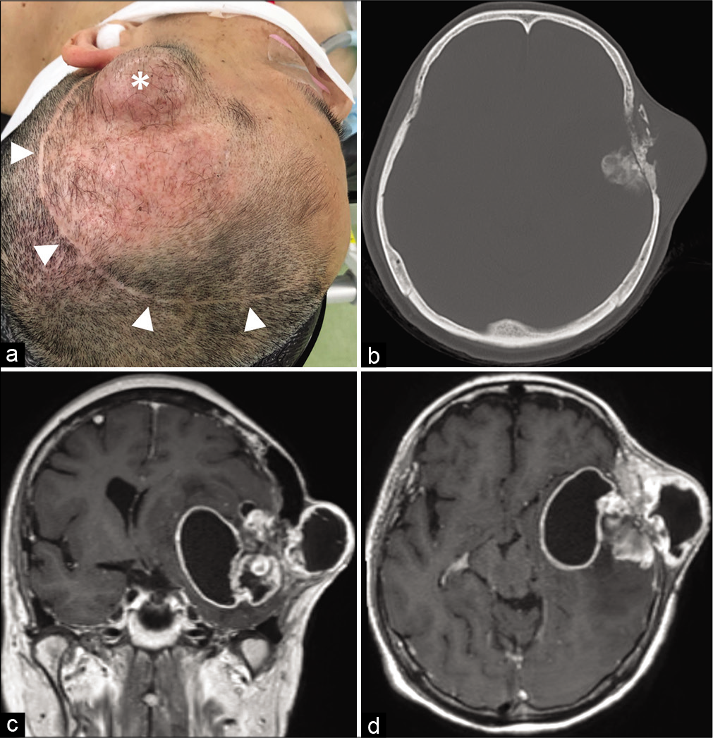
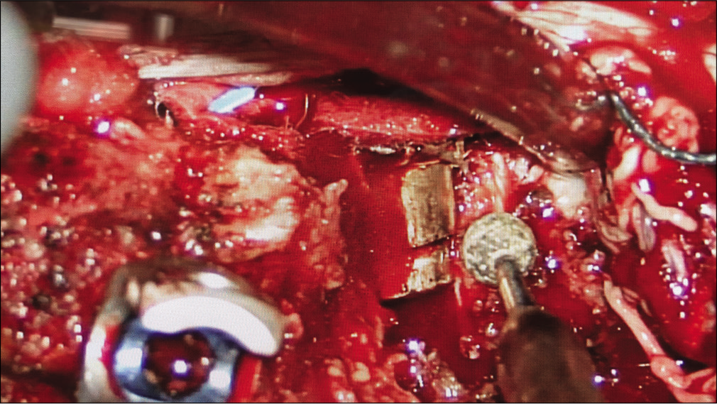
Background: Osteosarcoma (OS) is a malignant tumor of the bone, which rarely occurs in the head-and-neck regions as a primary or a secondary malignancy. Adequate surgical resection is currently the mainstay of treatment for head-and-neck OS; however, en bloc resection and reconstruction can be difficult because the anatomies of these regions are complex. We present a case of an OS arising from the temporal bone 40 years after radiation therapy, which was successfully treated with en bloc resection and a one-stage reconstruction using intraoperative tissue expansion technique.
Temporal muscle thickness and area are an independent prognostic factors in patients aged 75 or younger with aneurysmal subarachnoid hemorrhage treated by clipping
Date of publication: 14-Apr-2021
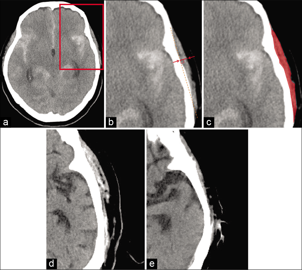
Background: Skeletal muscle mass is an important factor for various diseases’ outcomes. As for its indicators, temporal muscle thickness (TMT) and temporal muscle area (TMA) on the head computed tomography are useful, and TMT and TMA were reported as potential prognostic factors for aneurysmal subarachnoid hemorrhage (SAH). We examined the clinical characteristics, including TMT and TMA, of SAH patients aged 75 or younger.
Seizure in isolated brain cryptococcoma: Case report and review of the literature
Date of publication: 14-Apr-2021
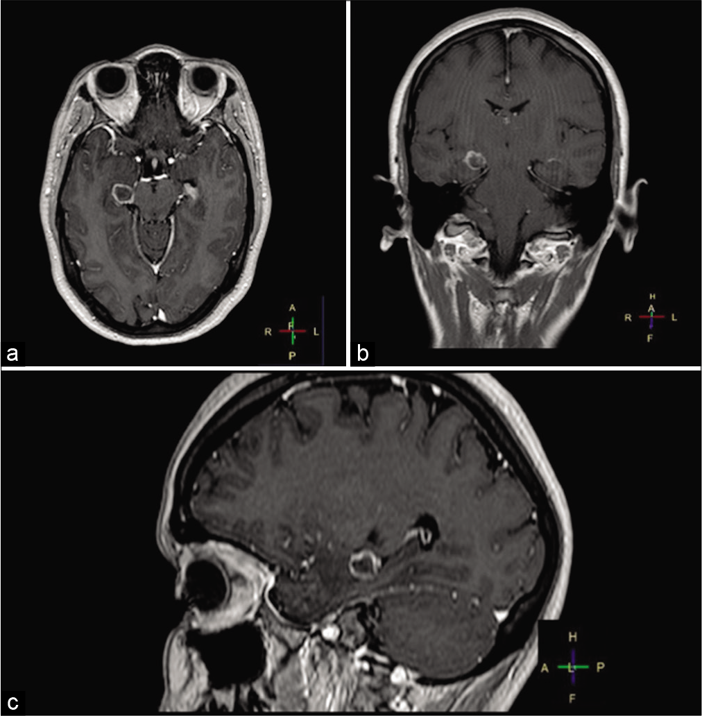
Background: Central nervous system (CNS) cryptococcosis is an invasive fungal infection predominantly seen among immunosuppressed patients causing meningitis or meningoencephalitis. Rarely, cryptococcosis can affect immunologically competent hosts with the formation of localized CNS granulomatous reaction, known as cryptococcoma. Common symptoms of CNS cryptococcoma are headaches, consciousness or mental changes, focal deficits, and cranial nerve dysfunction. Rarely, seizures are the only presenting symptom.
Cervical intradural intramedullary collision tumor of schwannoma and hemangioblastoma origin
Date of publication: 14-Apr-2021
Background: Primary spinal tumors are rare benign lesions that represent around 2–4% of all central nervous system neoplasms.[
Bullet retrieval from the cauda equina after penetrating spinal injury: A case report and review of the literature
Date of publication: 14-Apr-2021
Background: When gunshot injuries occur to the spine, bullet fragments may be retained within the spinal canal. Indications for bullet removal include incomplete spinal cord injury, progressive loss of neurologic function including injury to the cauda equina, and dural leaks with impending risk of meningitis.
Recurrent symptomatic vertebral hemangioma in pregnancy managed with decompression and vertebroplasty
Date of publication: 08-Apr-2021
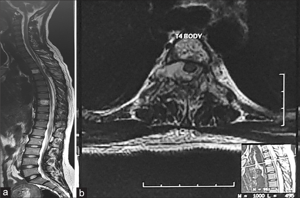
Background: Vertebral hemangioma is a benign vascular tumor of the spine that occurs in the endothelial lining of blood vessels. The majority of these lesions are detected incidentally on routine magnetic resonance imaging scans. Rarely, lesions can increase in size and result in neurological deterioration.
Extracranial vertebral artery to middle cerebral artery bypass in therapeutic internal carotid artery occlusion for epipharyngeal carcinoma: A technical case report
Date of publication: 08-Apr-2021
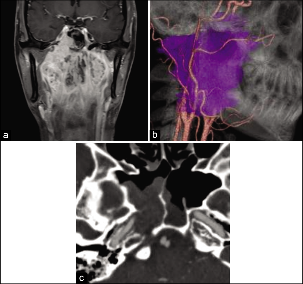
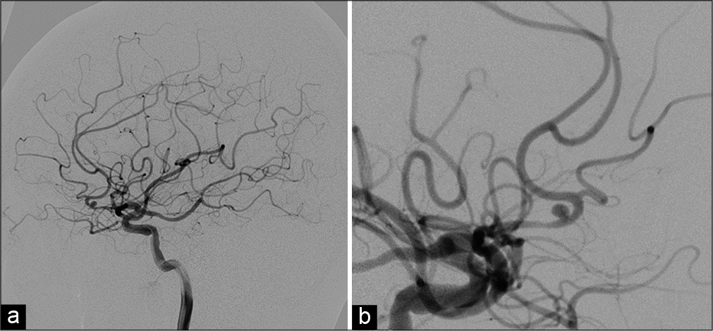
Background: Vertebral artery (VA) to middle cerebral artery (MCA) bypass is a rarely selected technique because a complex expanded dissection is required, and often, a better donor artery than VA exists. A good indication for VA-MCA bypass is the treatment of head-and-neck malignancies with the sacrifice of the internal carotid artery (ICA) or for carotid artery rupture.

Occipitofrontal switch for correction of anterior plagiocephaly planned through virtual mock surgery
Date of publication: 08-Apr-2021
Background: Unilateral coronal synostosis causing anterior plagiocephaly can result in restricted brain development and severe facial deformities. Various surgical procedures have been described for the correction of this deformity. Cranial vault remodeling, however, is associated with several complications. Occipitofrontal switching is a novel technique which utilizes a part of the contralateral occipital bone to reconstruct the frontal area. This is the first such case reported from India and first report where virtual mock surgery has been utilized for precision and improving outcome in this elegant procedure.
Coil embolization of subarachnoid hemorrhage with ruptured persistent primitive olfactory artery aneurysm
Date of publication: 08-Apr-2021
Background: Persistent primitive olfactory artery (PPOA) is a rare anomaly of the anterior cerebral artery. We experienced a rare case of subarachnoid hemorrhage caused by a ruptured saccular aneurysm of PPOA.

