

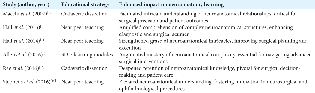
Enhancing neuro-ophthalmic surgical education: The role of neuroanatomy and 3D digital technologies – An overview
Date of publication: 29-Mar-2024
Background: Neuro-ophthalmology, bridging neurology and ophthalmology, highlights the nervous system’s crucial role in vision, encompassing afferent and efferent pathways. The evolution of this field has emphasized the importance of neuroanatomy for precise surgical interventions, presenting educational challenges in blending complex anatomical knowledge with surgical skills. This review examines the interplay between neuroanatomy and surgical practices in neuro-ophthalmology, aiming to identify educational gaps and suggest improvements.
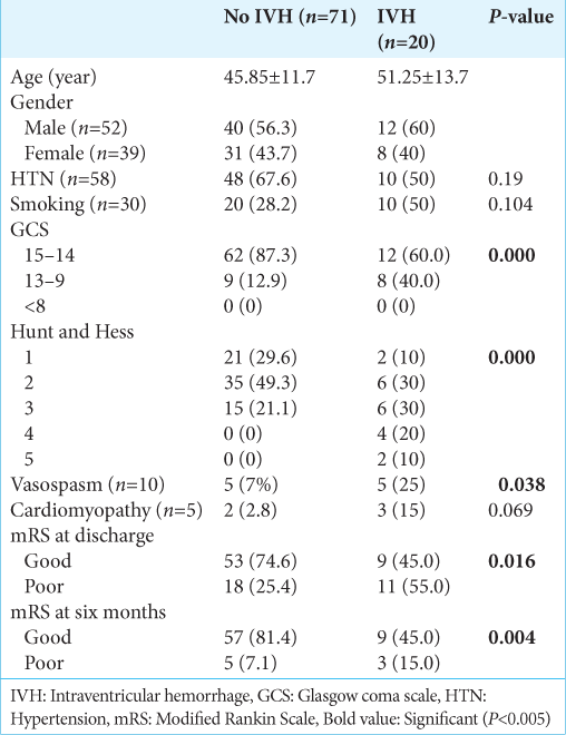
Analysis of the impact of intraventricular hemorrhage on the functional outcome of ruptured anterior cerebral artery aneurysm after clipping
Date of publication: 29-Mar-2024
Background: Various clinical symptoms and variables have been suggested as potential indicators of outcomes in patients with subarachnoid hemorrhage (SAH) resulting from ruptured intracranial aneurysms. The detailed discussion of the consequences of intraventricular hemorrhage (IVH), frequently reported in cases of anterior communicating artery (ACoA) aneurysms, is still pending. The study aimed to assess the results of aneurysm surgery performed early versus delayed in patients with SAH, specifically focusing on the occurrence of IVH.
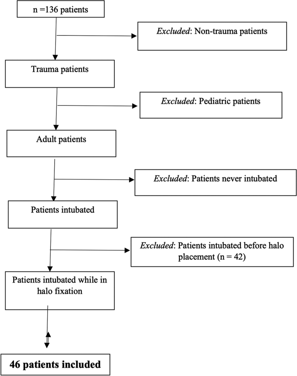
Airway risk associated with patients in halo fixation
Date of publication: 29-Mar-2024
Background: The halo fixation device introduces a significant obstacle for clinicians attempting to secure a definitive airway in trauma patients with cervical spine injuries. The authors sought to determine the airway-related mortality rate of adult trauma patients in halo fixation requiring endotracheal intubation.
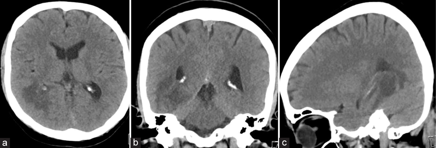
Occipital dermal sinus associated with infectious teratoma in an adult patient affected by Klippel–Feil syndrome: Rare case report and literature review
Date of publication: 22-Mar-2024
Background: The Klippel–Feil syndrome (KFS) is a rare congenital anomaly characterized by the fusion of cervical vertebrae, which may be associated with other malformations, such as dermoid tumors and teratoma. Some theories explain the embryology of these associations. Another condition that may be present is the dermal sinus (DS), communication between intracranial tumors and the subcutaneous tissue, and predisposing infections. This case report aims to describe an association between these three pathologies as well as correlate them from the literature. This report was based on medical records retrospectively reviewed associated with the systematic bibliographical consultation using indexed databases based on inclusion and exclusion methods.
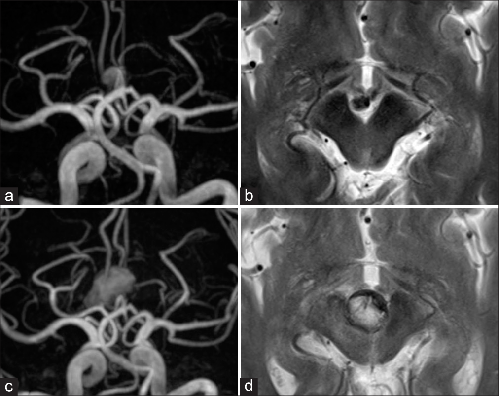
Successful shrinkage of a recurrent partially thrombosed symptomatic large basilar tip aneurysm using a Target 3D Coil
Date of publication: 22-Mar-2024
Background: Standalone coil embolization is often less effective for partially thrombosed intracerebral aneurysms (PTIA) because of the risk of frequent recurrence if the coil migrates into the thrombus. This report describes a case of PTIA at the basilar tip in which simple coil embolization using a Target 3D Coil resulted in sustained remission without recurrence during long-term follow-up.
On the balance beam: facing the challenges of neurosurgical education in the third millennium
Date of publication: 22-Mar-2024
Background: Neurosurgery is one of the most complex and challenging areas of medicine, and it requires an ongoing commitment to education and expertise. Preparing young neurosurgeons with comprehensive education that can allow them to achieve high professional standards is a pivotal aspect of our profession.
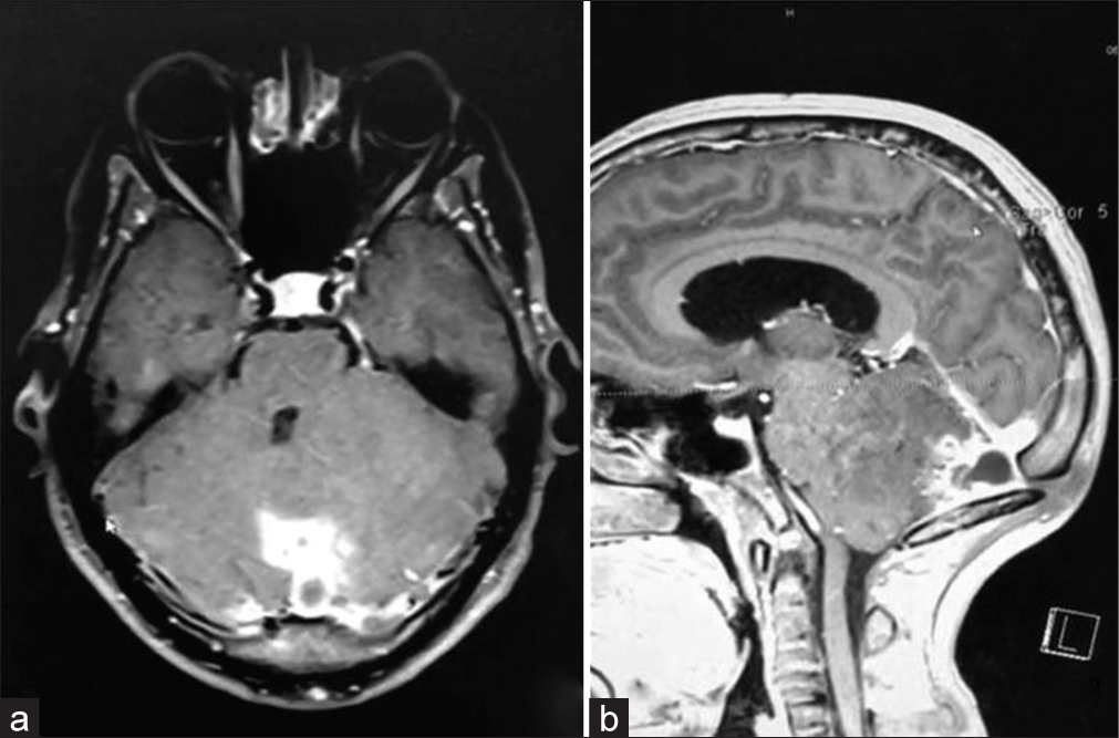
Intracranial malignant peripheral nerve sheath tumor: A case report and comprehensive literature review
Date of publication: 22-Mar-2024
Background: Malignant peripheral nerve sheath tumors (MPNSTs) are rare malignant soft-tissue sarcomas arising from peripheral nerves. Little data exist regarding MPNST originating intracranially. Here, we present a 7th/8th nerve complex MPNST, discuss the treatment strategy and patient outcome, and provide a comprehensive review of existing literature.
Secondary normal pressure hydrocephalus following pituitary apoplexy: A case report
Date of publication: 22-Mar-2024
Background: Although secondary normal pressure hydrocephalus (sNPH) can occur in various central nervous system diseases, there are no reports of sNPH caused by pituitary lesions. Herein, we present a unique case of sNPH caused by pituitary apoplexy.
A case report of an unusual cerebral hydatid cyst
Date of publication: 22-Mar-2024
Background: Intracranial hydatid cyst is an exceedingly uncommon condition. Typically, it manifests as hydatid cysts in the liver, lungs, kidney, and spleen. In this report, we present a rare case of a hydatid cyst located in the brain, exhibiting atypical radiological characteristics, and successfully treated with complete microsurgical excision.

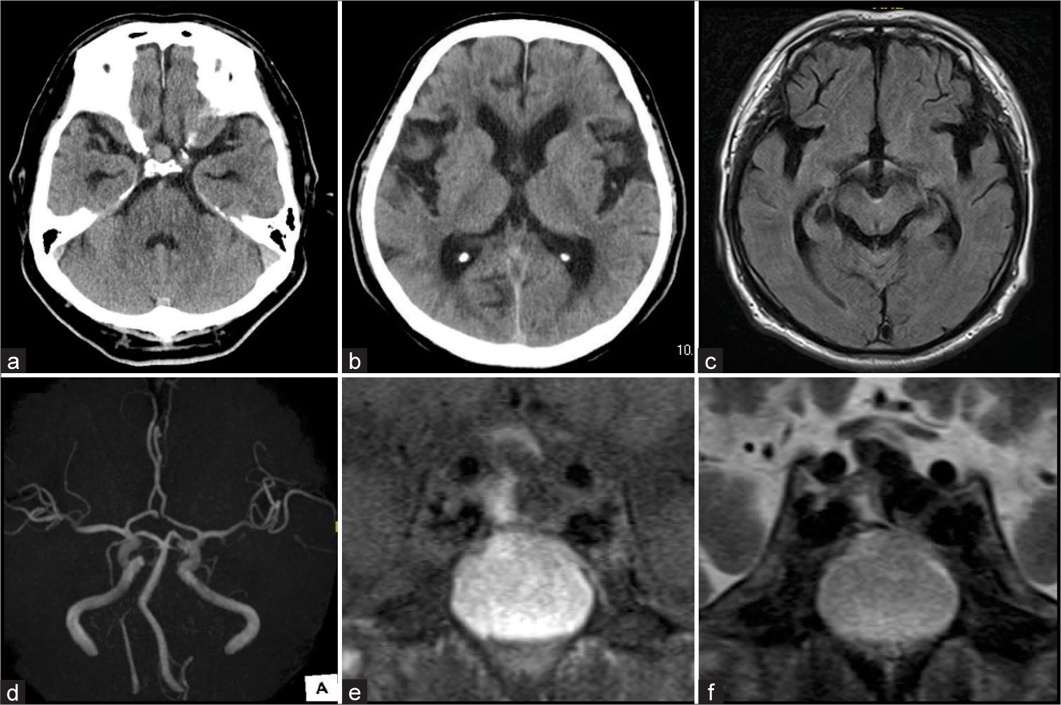
Spontaneous thrombosis and calcification of giant cavernous carotid artery aneurysm: A rare case and management insights
Date of publication: 22-Mar-2024
Background: Giant cavernous carotid artery aneurysms (>25 mm) are rare (3–5%), with some prone to spontaneous thrombosis (10–20% complete). We present a unique case of one of the largest aneurysms spontaneously thrombosing and calcifying.

