

An uncommon intramedullary tumor: Primary medullary cone melanoma
Date of publication: 18-Jul-2020
Background: Melanoma is the third most common primary tumor to metastasize to the central nervous system (CNS). However, primary CNS melanoma is very rare, and primary intramedullary melanoma is even less frequently encountered, with only 27 cases published in the literature. There are no pathognomonic imaging characteristics, therefore, the diagnosis must be confirmed immunohistologically and the preferred treatment is the gross total resection.
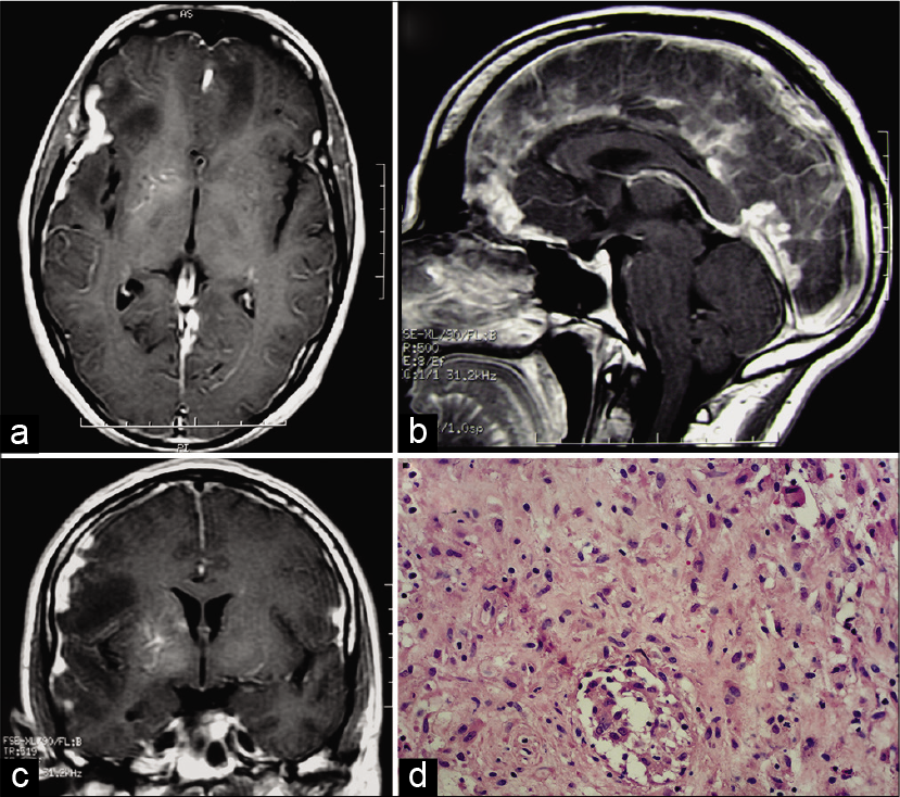
Hypertrophic cranial pachymeningitis coinfection with tuberculosis and actinomycosis
Date of publication: 18-Jul-2020
Abstract
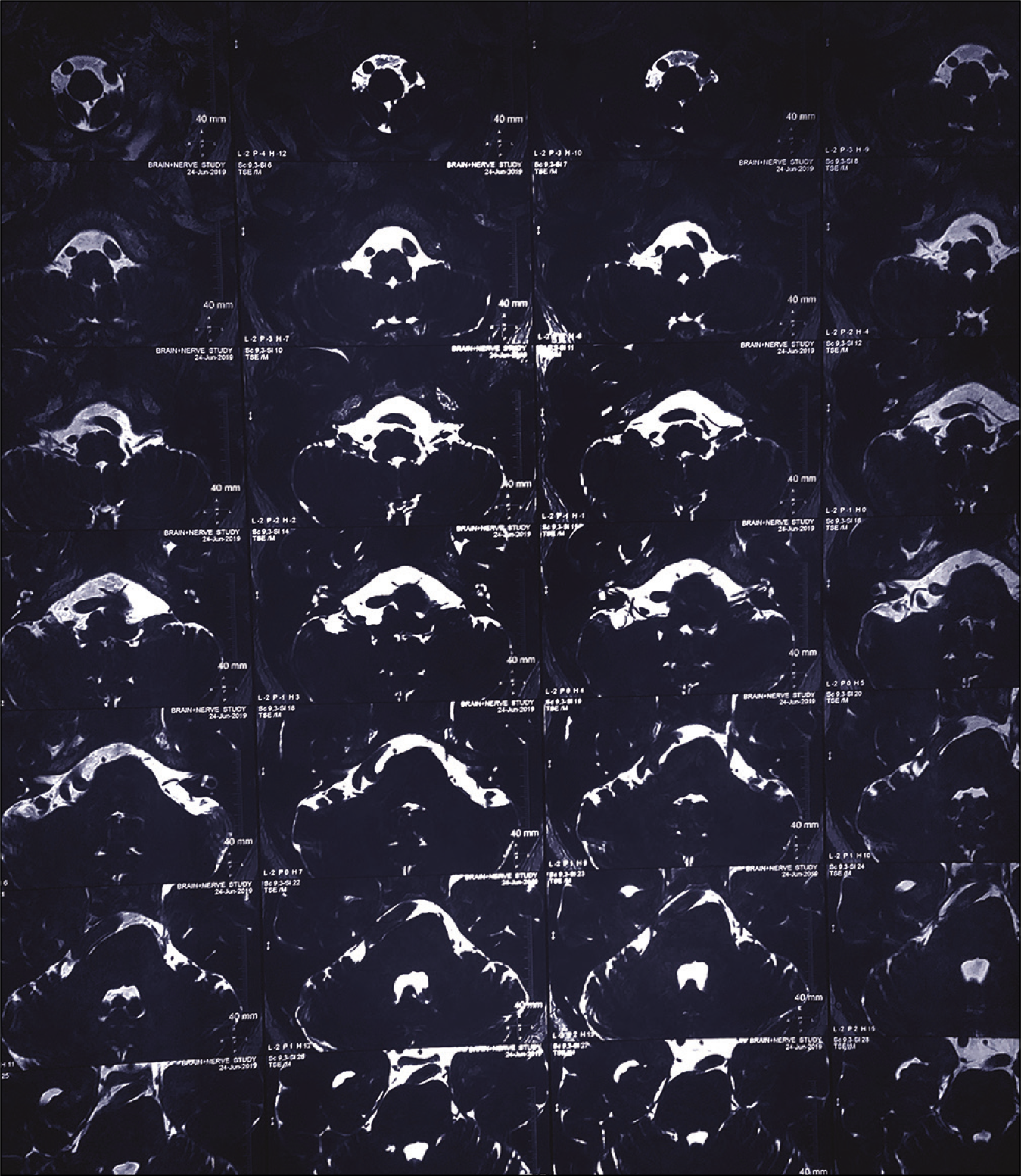
This is a rare case report about hypertrophic cranial pachymeningitis coinfection with tuberculosis and actinomycosis in a 35-year-old male. The patient presented with progressive headache, paraesthesia, and blurred vision. Dural biopsy, histology, and cultures are imperative in pachymeningitis for establishing the diagnosis and guiding treatment.
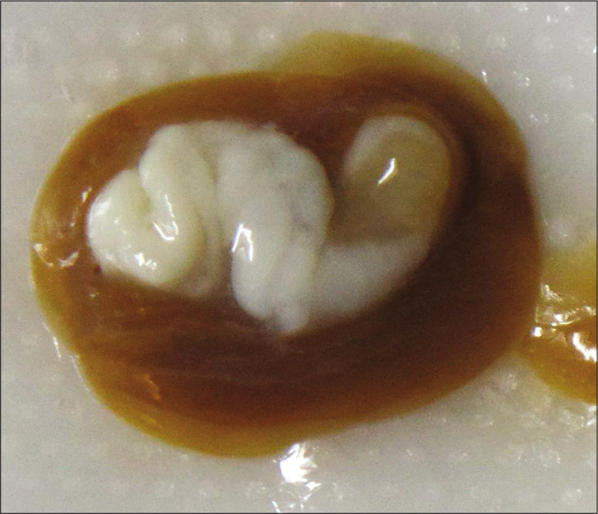
Intraventricular adult Taenia solium causing hydrocephalus: A case report
Date of publication: 18-Jul-2020
Background: Neurocysticercosis (NCC) is the most common parasitic infection of the central nervous system worldwide and is caused by the larval form of the tapeworm Taenia solium. In general, T. solium larval form may be located in the neuraxis, resulting in pathology. Here, we report a rare case of female with a history of adult onset seizures presenting with adult form T. solium in the fourth ventricle, causing hydrocephalus.
Posttraumatic thoracic epidural capillary hemangioma – A rare case report
Date of publication: 11-Jul-2020
Background: Capillary hemangiomas are benign vascular lesions commonly seen in subcutaneous tissues. The most common site of origin is from the vertebral body, and only a few cases of isolated lesions in thoracic epidural space, especially after trauma, have been reported in the literature.
Hemifacial spasm due to multiple neurovascular conflicts: A conundrum
Date of publication: 11-Jul-2020
Aggressive internal and external decompression as a life-saving surgery in a deeply comatose patient with fixed dilated pupils after severe traumatic brain injury: A case report
Date of publication: 11-Jul-2020
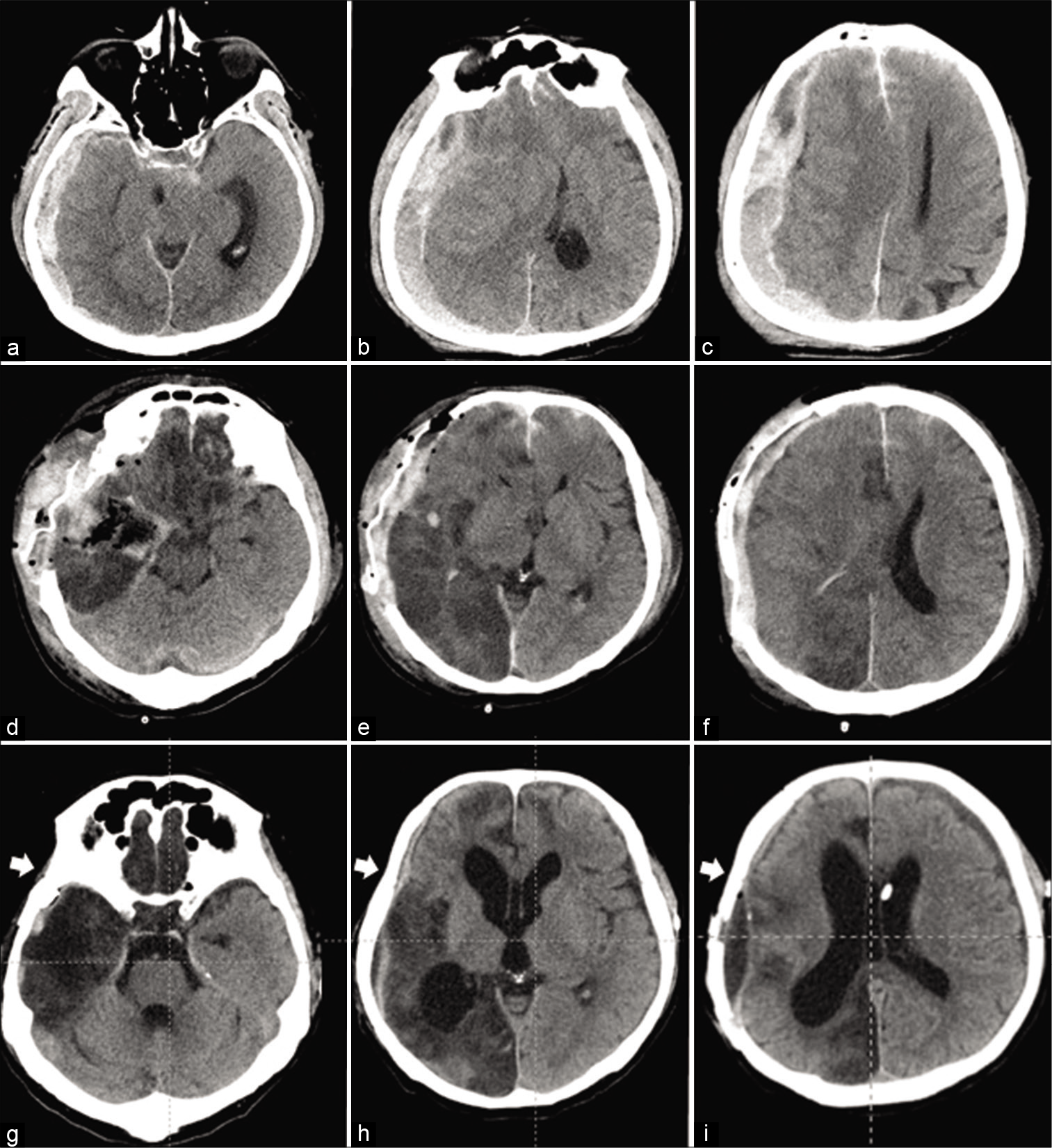
Background: To maximize control of the intracranial pressure in deeply comatose patients with malignant cerebral swelling, combination of the surgical techniques for internal and external brain decompression may be reasonable, as demonstrated in the presented case.
Extremely rare case of retropharyngeal space benign plexiform schwannoma - Excised through Smith- Robinson Approach
Date of publication: 11-Jul-2020
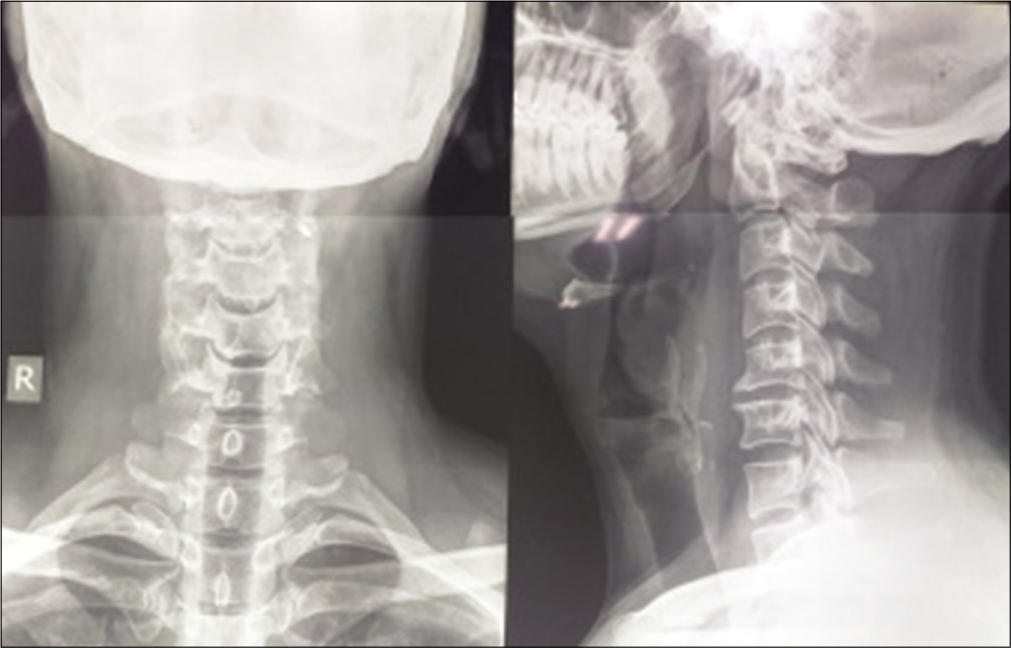
Background: Approximately 25–45% of schwannomas are typically slow-growing, encapsulated, and noninvasive tumors that occur in the head-and-neck region where they rarely involve the retropharyngeal space. Here, we report deep-seated benign plexiform schwannoma located in the retropharyngeal C2-C5 region excised utilizing the Smith-Robinson approach.

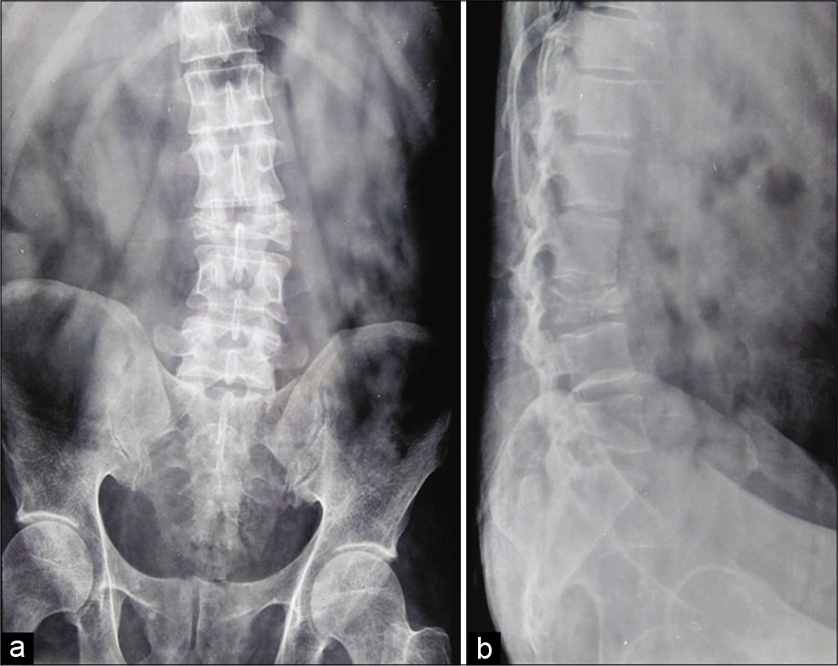
Extensive vertebral scalloping in a thoracolumbar junction spinal schwannoma
Date of publication: 11-Jul-2020
Abstract
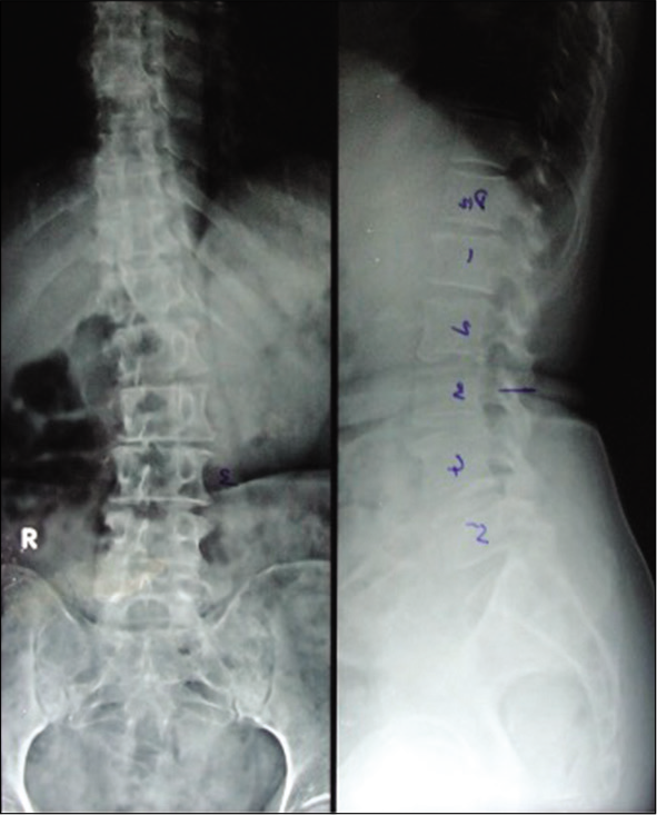
Severe vertebral scalloping in spinal schwannoma is very rare. When present, extensive scalloping of the vertebral bodies possesses significant treatment challenges in patients with spinal tumors. We present the computed tomography scan and magnetic resonance images of spinal schwannoma with marked vertebral scalloping in a 40-year-old Nigerian.
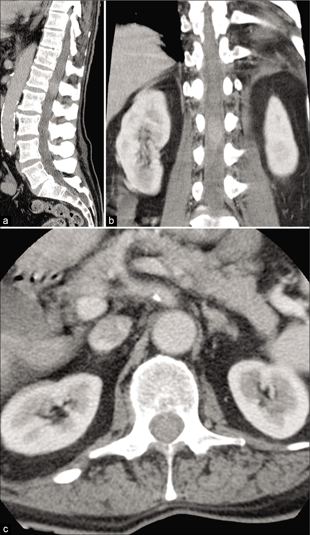
Salmonella Typhi dorsolumbar spondylodiscitis mimicking tuberculosis – An interesting case report
Date of publication: 11-Jul-2020
Background: Salmonella rarely causes spinal infections in patients other than those who are immunocompromised or have sickle cell anemia. Further, most cases occurring in healthy individuals have preexisting gastrointestinal infections. Here, we present a case of pyogenic spondylodiscitis attributed to Salmonella Typhi, in an immunologically normal patient without gastrointestinal pathology.
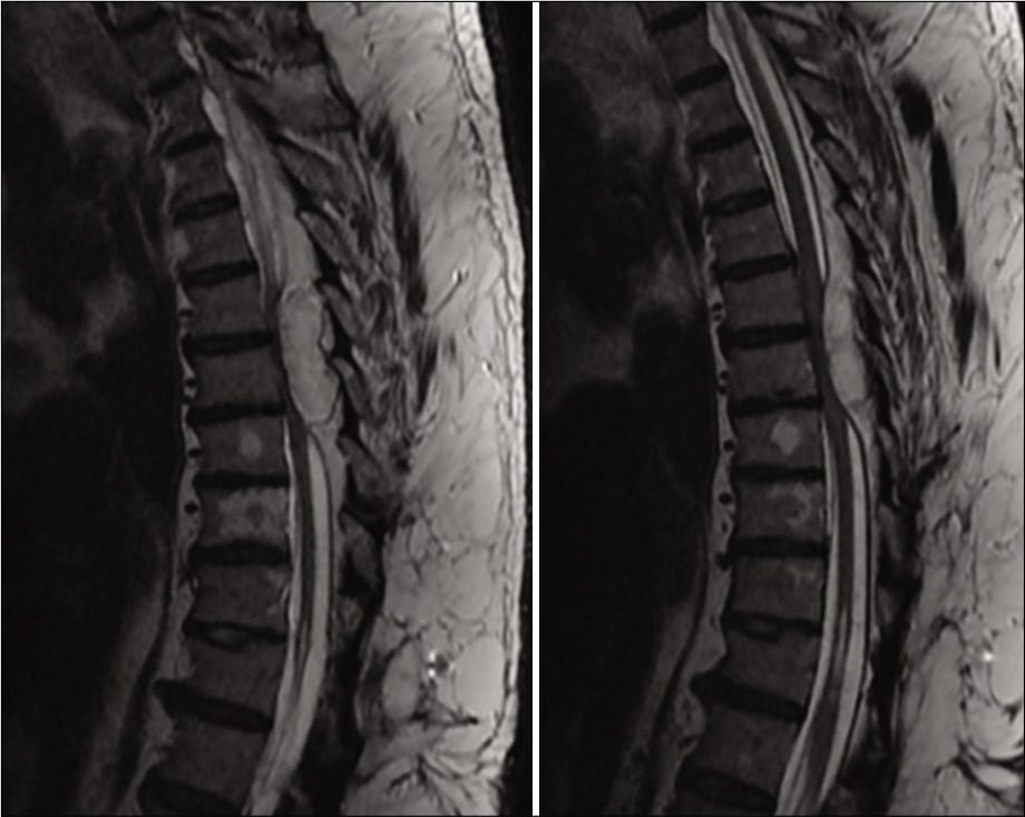
Metastatic small cell neuroendocrine tumor with spinal cord compression – Understanding a rare entity
Date of publication: 11-Jul-2020
Background: Metastatic spinal cord compression with carcinoid tumor as primary is a rare entity with its own diagnostic dilemmas and surgical challenges. Most of these neuroendocrine tumors arise from the gastrointestinal tract or lungs with metastasis to spine in <2% cases. Early diagnosis in an orderly manner is of significance as most of it is delayed due to slowly developing symptoms. Furthermore, prompt management has been an important factor as morbidity and mortality are high in such cases and surgical intervention if needed, which can be a challenge due to disturbed alignment, complex regional anatomy, and careful handling of spinal cord.

