

Intramedullary craniovertebral junction metastasis leading to the diagnosis of underlying renal cell carcinoma
Date of publication: 13-Jun-2020
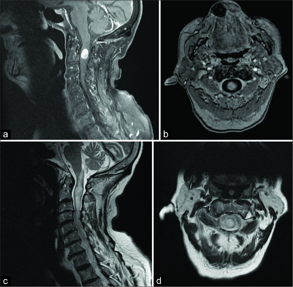
Background: Intramedullary spinal cord metastases represent 4–8.5% of the central nervous system metastases and affect only 0.1–0.4% of all patients. Those originating from renal cell carcinoma (RCC) are extremely rare. Of the eight patients described in the literature with metastatic RCC and intramedullary cord lesion, only five were found in the cervical spine. Here, the authors add a 6th case involving an RCC intramedullary metastasis at the C1–C2 level.
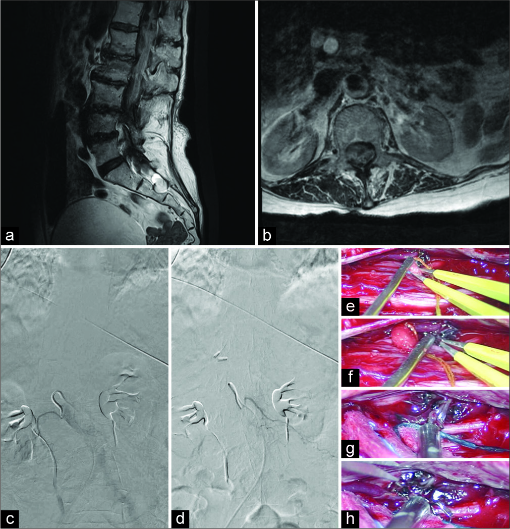
Unique Bone Suture Anchor Repair of Complex Lumbar Cerebrospinal Fluid Fistulas
Date of publication: 13-Jun-2020
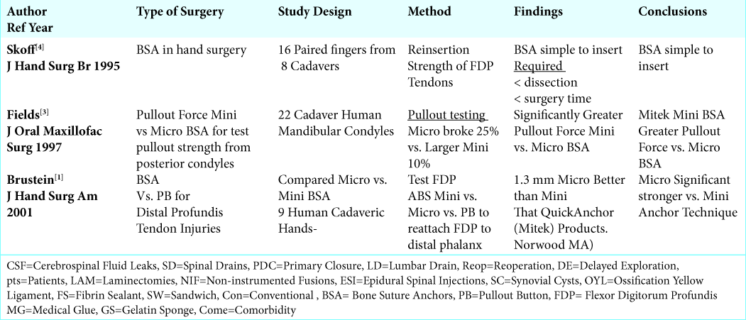
Background: Spine surgeons encounter occasional complex cerebrospinal fluid fistulas/dural tears (CSF/DT) during lumbar spinal surgery. In some cases, these leaks are found during the index procedure, but others may appear postoperatively, or in the course of successive procedures. Here we asked, whether these complex CSF fistulas/DT could be more readily repaired utilizing a “bone suture anchor” technique, particularly where there is no residual dural margin/remnant.
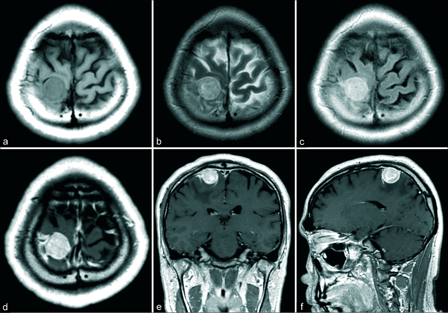
Supratentorial intraventricular rosette-forming glioneuronal tumors – Case report and review of treatment paradigms
Date of publication: 06-Jun-2020
Background: Rosette-forming glioneuronal tumors (RGNT) are slow-growing WHO Grade I tumors that are characterized by mixed histology and rosette formation. Although typically located in the posterior fossa, these tumors can rarely originate elsewhere. Here, we describe the fourth case in literature where an RGNT was localized to the lateral ventricles and detail the treatment approach.
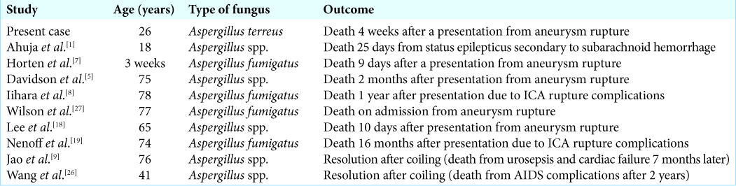
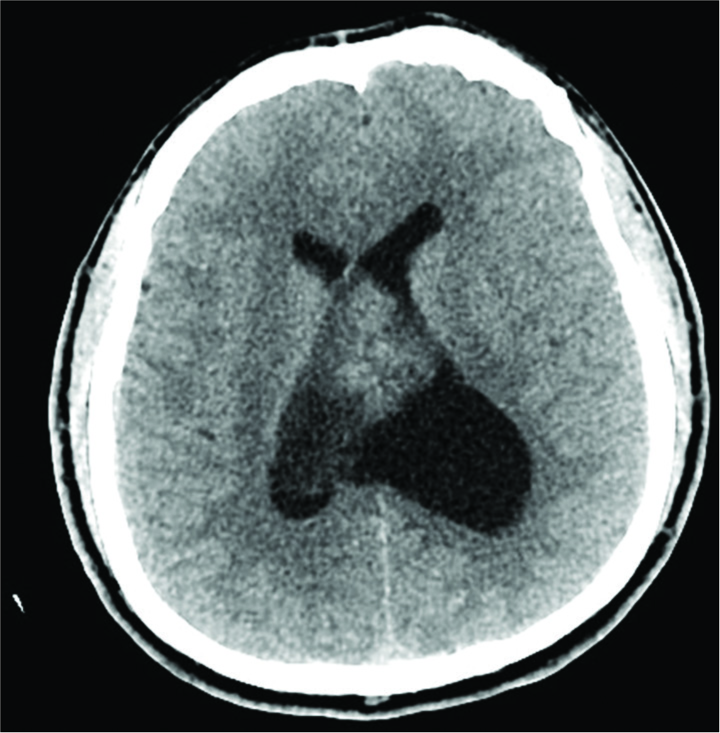
Fungal mycotic aneurysm in a patient with Aspergillus terreus chronic meningoencephalitis
Date of publication: 06-Jun-2020
Background: Central nervous system involvement due to aspergillosis is an extremely serious entity, particularly in patients with severe neutropenia, hematological diseases, or post-transplant cases. Immunocompetent patients can be infected by intense exposure, particularly iatrogenic after invasive procedures.
Parvimonas micra: A potential causative pathogen to consider when diagnosing odontogenic brain abscesses
Date of publication: 06-Jun-2020
Background: Brain abscess is a life-threatening entity which requires prompt and long-term antibiotic therapy, generally associated with surgical drainage, and eradicating the primary source of infection. Parvimonas micra (Pm) has only been reported once before as the lone infecting organism of an orally originated, solitary brain abscess. Diagnosing brain abscesses caused by this Gram-positive anaerobic coccus, constituent of the oral cavity flora, is challenging, and an optimal treatment regimen has not been well established. We report the diagnosis and successful treatment of a Pm caused odontogenic brain abscess.
Hemifacial spasm caused by the brainstem developmental venous anomaly: A case report and review of the literature
Date of publication: 06-Jun-2020
Background: Hemifacial spasm (HFS) is usually caused by vascular compression of the root exit zone (REZ) of the facial nerve. Dual compression of the REZ by veins and arteries is also associated with HFS, but venous origin alone is rarely reported. We present a rare case of HFS caused by the brainstem developmental venous anomaly (DVA) treated with microvascular decompression (MVD).
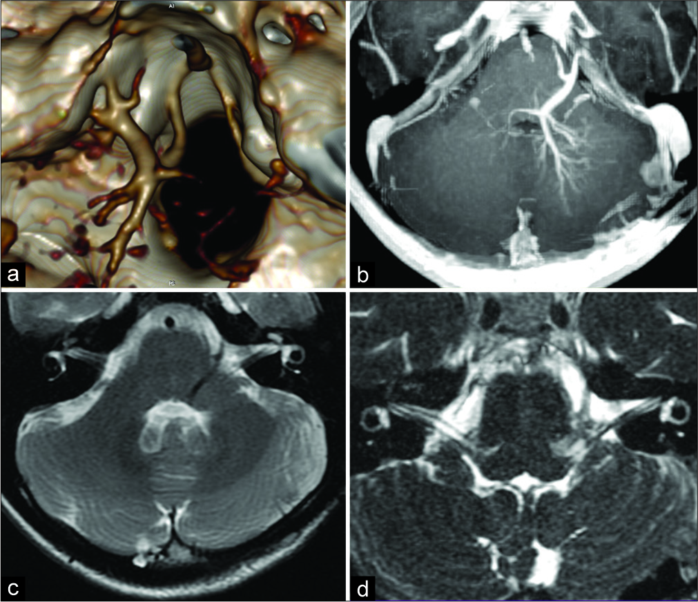
Spinal dural arteriovenous fistula masquerading as subdural hematoma
Date of publication: 06-Jun-2020
Background: This case highlights an angiographically occult spinal dural AVF presenting with a spinal subdural hematoma. While rare, it is important that clinicians be aware of this potential etiology of subdural hematomas before evacuation.
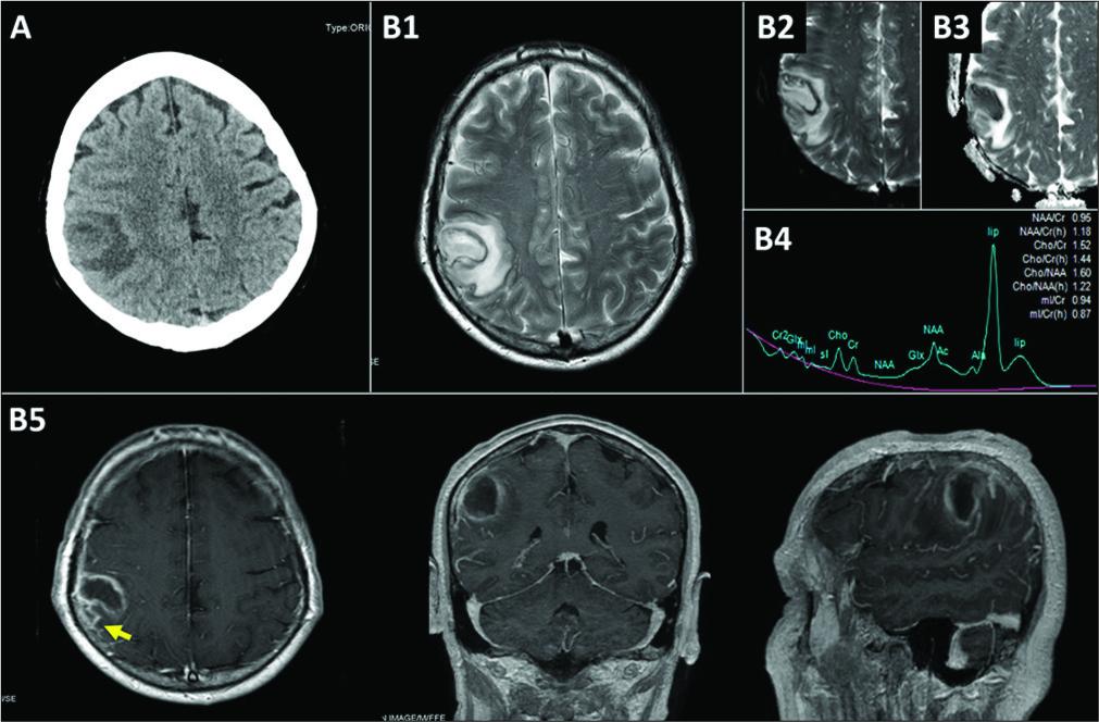
Supratentorial convexity schwannoma unrelated to cranial nerves: Case report and review of the literature
Date of publication: 06-Jun-2020
Background: Intracranial schwannoma not related to cranial nerves is rare entity, and difficult to be diagnosed preoperatively. Here, we experienced a case of convexity schwannoma mimicking convexity meningioma, and discuss about the characteristics of such cases based on the past published reports.
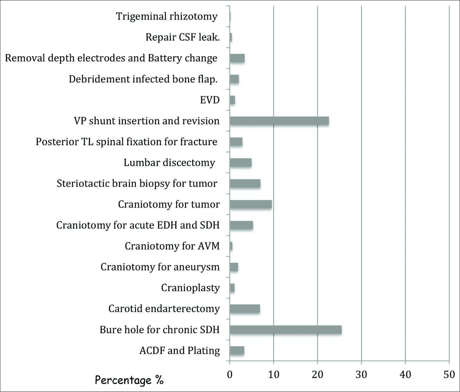
Effect of length time to surgery on postoperative hospital length of stay among neurosurgical patients
Date of publication: 06-Jun-2020
Background: In most hospitals, inpatient urgent surgery is triaged based on the degree of urgency and time of surgical booking. A longer wait for semi-urgent surgery due to sharing resources between specialties might impact the postoperative course. The objective of this study is to determine the effect of length time to semi- urgent surgery on postoperative hospital length of stay among neurosurgical patients.
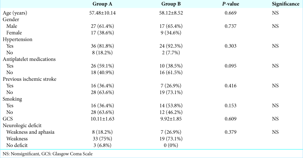
Early versus delayed surgical evacuation of spontaneous supratentorial intracerebral hematoma: A prospective cohort study
Date of publication: 06-Jun-2020
Background: The optimum timing for surgical evacuation of spontaneous supratentorial intracerebral hematoma (ICH) is still controversial. The aim of this study was to compare the clinical outcome following early versus delayed surgical evacuation of spontaneous supratentorial ICH.

