

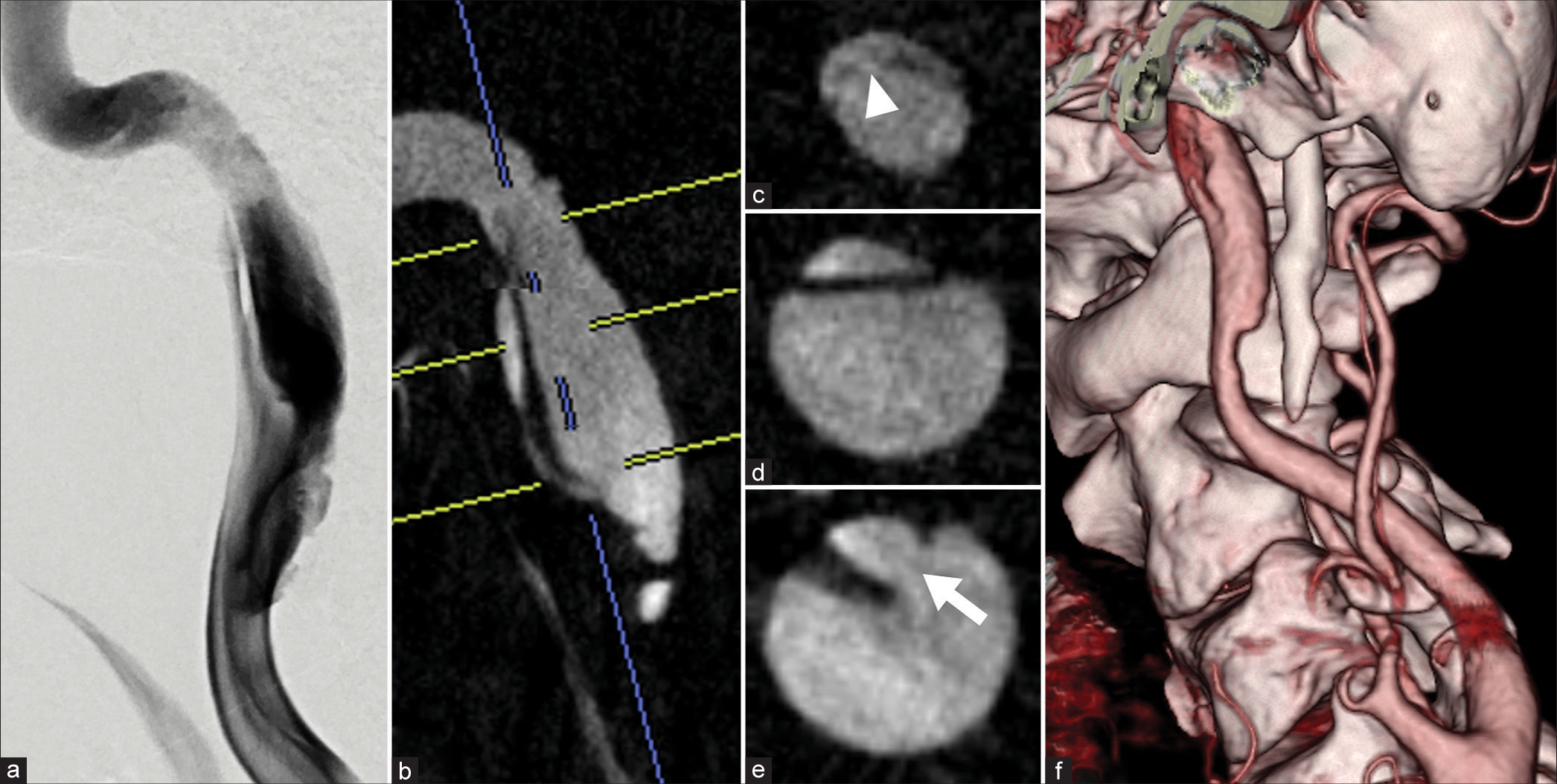
Extracranial internal carotid artery-dissecting aneurysm having a re-entry tear and causing lower cranial nerve palsies treated with flow-diverting stent: A case report
Date of publication: 12-Apr-2024
Background: Extracranial internal carotid artery (ICA)-dissecting aneurysms (DAs) rarely cause re-entry tears and lower cranial nerve palsies. The therapeutic strategies for these pathologies are not well established. This report presents a case of an extracranial ICA -DA with a re-entry tear that caused lower cranial nerve palsy.
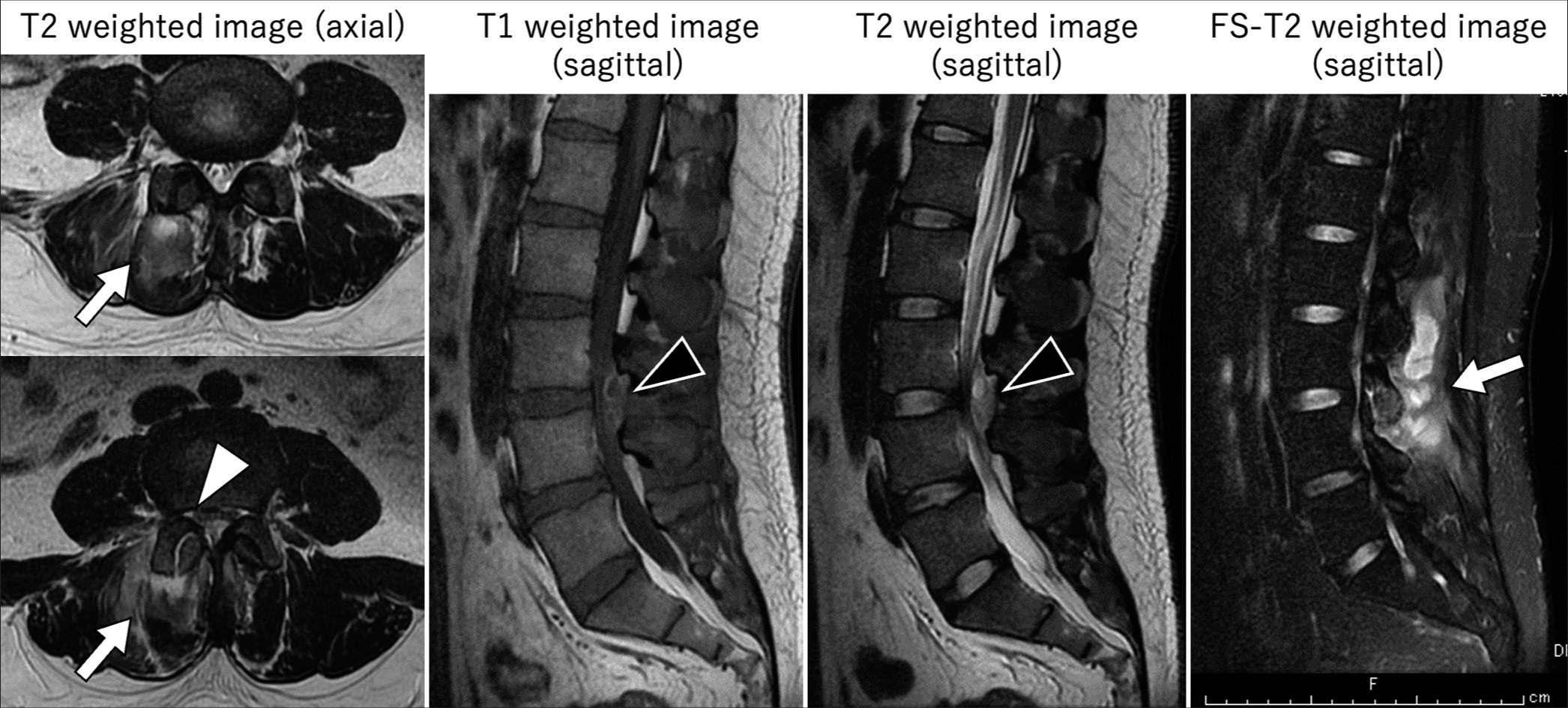
Psoriasis vulgaris of the skin caused a L3-L4 lumbar epidural spinal abscess
Date of publication: 12-Apr-2024
Background: In a 31-year-old male, psoriasis vulgaris (PV) of the skin caused paraparesis attributed to a L3-L4 epidural spinal abscess that required emergent surgical decompression.
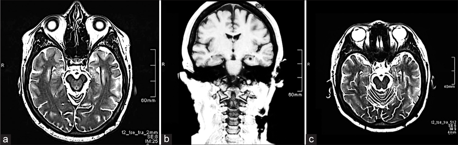
Tuberculoma mimicking en-plaque meningioma in a 45-year-old male: A case report
Date of publication: 05-Apr-2024
Background: Tuberculoma mimicking en-plaque meningioma is a rare variant of tuberculoma. A few cases were reported in the literature. The radiological appearance can be mistakenly diagnosed as en-plaque meningioma.
Adequate control of seizures in a case of lead migration and neuromodulation of the posterior Sylvian junction: A case report
Date of publication: 05-Apr-2024
Background: This report aims to describe the neuromodulation effect on seizure control in a patient with a left hippocampal migrated electrode to the Posterior Sylvian Junction (PSJ) during a follow-up of 17 years.
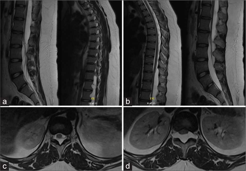
Extradural spinal cyst in a pediatric patient: A case report
Date of publication: 05-Apr-2024
Background: Spinal extradural arachnoid cysts comprise <1% of all spinal lesions and are rare findings in pediatric patients. The pathogenesis of spinal extradural arachnoid cysts is not well known but is thought to most commonly be due to congenital dural defects. Other origins include trauma, inflammation, or infection, such as arachnoiditis. Spinal magnetic resonance imaging is the gold standard for diagnosis, showing a fluid-filled space dorsal to the spinal cord with signal intensity akin to cerebrospinal fluid (CSF) and often the site of dural defect with CSF leak. While most spinal extradural arachnoid cysts are asymptomatic, large cysts can compress the spinal cord or nerve roots, leading to myelopathy, radiculopathy, or focal pain symptoms. In such cases, surgical management is indicated.
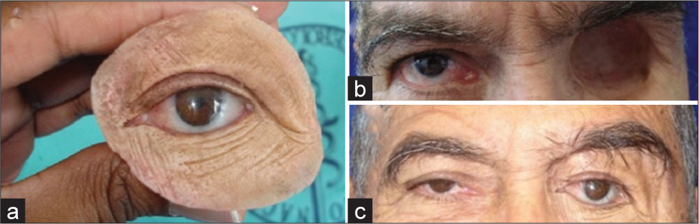
Surgical treatment of orbital tumors in a single center: Analysis and results
Date of publication: 05-Apr-2024
Background: Orbital tumors, arising within the bony orbit and its contents, present diverse challenges due to their varied origins and complex anatomical context. These tumors, classified as primary, secondary, or metastatic, are further subdivided into intraconal and extraconal based on their relationship with the muscle cone. This classification significantly influences surgical approach and management. This study highlights surgical experiences with orbital tumors, underscoring the importance of tailored surgical approaches based on the lesion’s site and its proximity to the optic nerve.
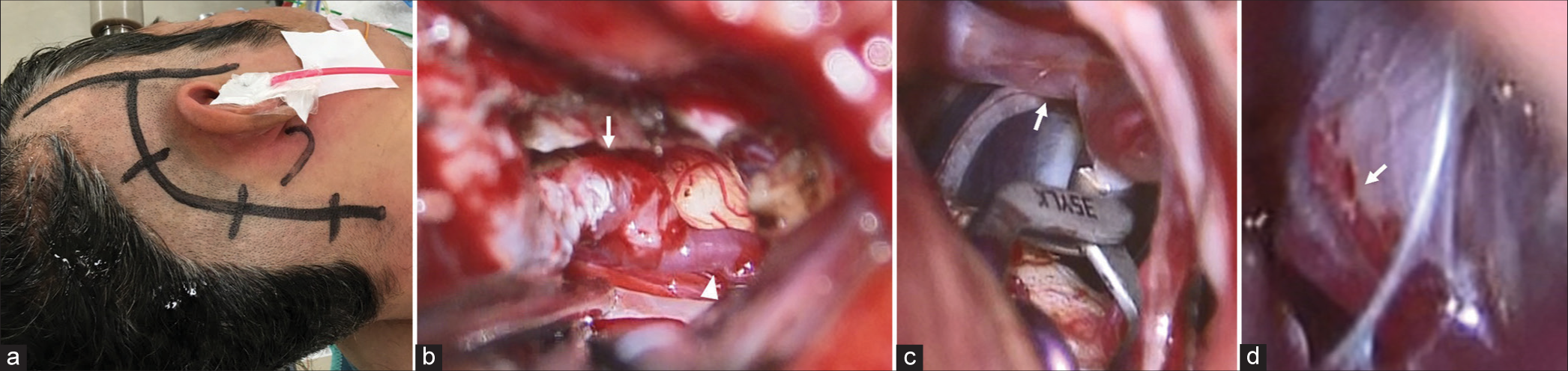
A case of bilateral vertebral artery dissection treated by bilateral surgical occlusion and low-flow bypass
Date of publication: 05-Apr-2024
Background: Bilateral vertebral artery dissection aneurysm (VADA) is a rare condition that leads to severe stroke. However, the surgical strategy for its treatment is controversial because the pathology is very complicated and varies in each case. Here, we report a case of bilateral VADA that was successfully treated with staged bilateral VADA occlusion and low-flow bypass.
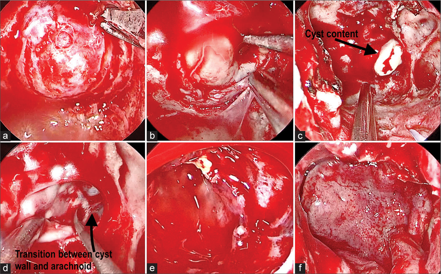
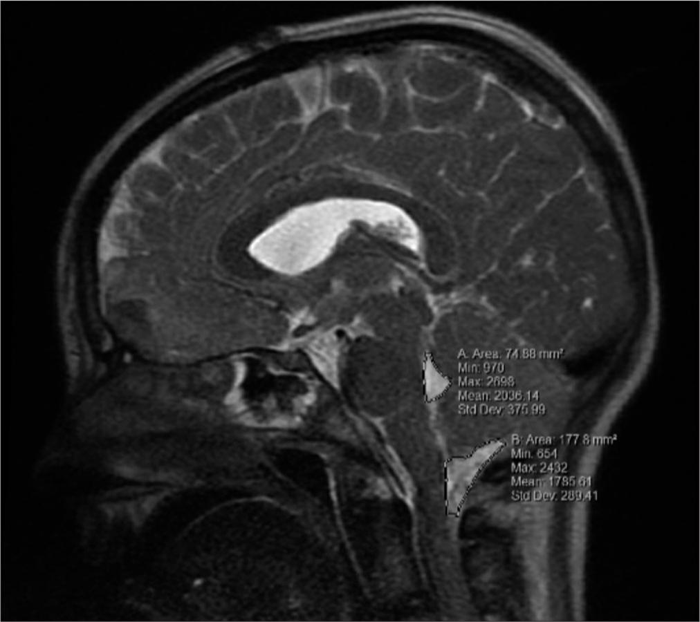
Accurate preoperative diagnosis of a Rathke cleft cyst with the aid of a novel classification for sellar cystic lesions and a diagnostic algorithm decision: Tools for differentiating cystic sellar lesions with a representative case
Date of publication: 05-Apr-2024
Background: Rathke’s cleft cyst (RCC) is a benign lesion in the sellar and suprasellar compartments. Similarly, pituitary adenomas can present with cystic morphology, making it a differential diagnosis when evaluating a patient with a cystic lesion in the sellar region. Surgical goals differ between RCCs and pituitary adenomas as the first can achieve remission of symptoms with cyst decompression in contrast to pituitary adenomas where complete resection would be the main goal. Imaging analysis alone may not be sufficient to define a preoperative surgical plan. The combination of imaging and conjoined use of validated tools may provide valuable insights to the clinician when defining a surgical approach.
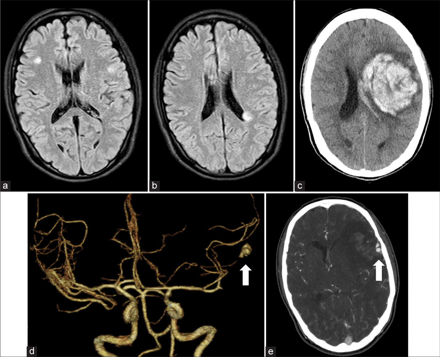
Intracranial mycotic aneurysm rupture following cupping therapy
Date of publication: 05-Apr-2024
Background: Cupping therapy is an alternative treatment that uses a small glass cup to suck the skin with a needle and has been used to manage skin problems and pain. However, serious complications have been reported. Herein, we describe a case of intracranial mycotic aneurysm rupture after cupping therapy.
Prognostic and morphological factors in pediatric cerebellar contusions
Date of publication: 05-Apr-2024
Background: Although uncommon, cerebellar contusions are associated with significant morbidity and mortality. Literature is lacking in the prognostic and morphological factors relating to their clinical picture and outcomes, especially within children. The objective of this study is to evaluate prognostic and anatomic factors in the clinical picture of cerebellar contusions, including effacement of the 4th ventricle and cisterna magna.

